AI-powered image analysis
As live-cell imaging experiments scale, image analysis can become a major bottleneck. Conventional threshold- and intensity-based methods often require extensive parameter tuning and can struggle with challenging cell types, variable morphologies, and label-free imaging, making analysis time-consuming and subjective.
AI-powered image analysis offers a more robust approach, improving cell segmentation across diverse imaging conditions while reducing manual optimization and user-to-user variability.
In this issue of TekTalk, we highlight two Agilent solutions that bring these capabilities to live-cell analysis. Both integrate powerful AI models directly within the analysis software, enabling local processing without the need to transfer image data off premises. Agilent xCELLigence RTCA eSight AI software enhances label-free cell segmentation to quantify cell count, confluence, proliferation, and treatment response alongside real-time impedance measurements. Agilent BioTek Gen5 AI cell identification module provides accurate label-free and fluorescent label-based segmentation for quantifying cell numbers and boundaries, while revealing subtle morphological changes associated with cellular response, health, and phenotype (Figure 1).
Together, these AI-powered tools provide a simpler, less subjective approach to image analysis, reducing manual optimization, and accelerating the path from image acquisition to reliable, biologically meaningful results.
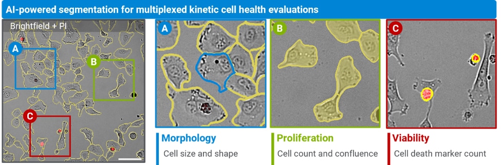
Figure 1. AI-powered segmentation supports quantitative assessment of multiple cell health parameters, including morphology, proliferation, and viability.
Featured Application Notes
AI-Powered, Label-Free Cell Segmentation, Counting, and Fluorescence-Based Classification
Accurate and reproducible cell segmentation is critical for extracting biological insights from microscopy images. The AI-powered, label-free cell segmentation feature in the Agilent xCELLigence RTCA eSight system uses deep learning to automatically segment single cells from brightfield images without fluorescent labeling or manual optimization. It supports accurate cell counting, proliferation analysis, fluorescence-based subpopulation classification, and high-throughput 384-well screening. Delivering results comparable to fluorescence-based methods, this noninvasive solution streamlines image analysis, improves reproducibility, and enhances confidence in cell-based research and drug discovery.
AI‑Enabled Cell Morphology Profiling Using Automated Imaging and Analysis
AI-enabled cell identification can make epithelial‑to‑mesenchymal transition (EMT) imaging workflows more quantitative and reproducible. In dense A549 monolayers, conventional intensity-based segmentation struggles to define cell boundaries, especially after TGF-β-induced cell elongation. The Gen5 AI cell identification module supported more robust whole-cell segmentation from DAPI and phalloidin images, enabling morphology metrics that tracked with established EMT marker responses and linked molecular changes to shifts in cell shape and cytoskeletal organization.
Tek Tips
Choose the Analysis Method That Fits the Application
AI-based segmentation is particularly valuable for label-free imaging, complex or changing cell morphologies, and workflows where conventional thresholding requires significant parameter optimization. AI can provide more consistent segmentation across these challenging conditions with less manual tuning.
For simpler applications with strong, well-defined fluorescence signals, conventional analysis may still be the most efficient choice. For example, threshold-based identification of fluorescently labeled nuclei can provide a fast, straightforward approach for cell counting when segmentation requirements are simple. The AI-Powered, Label-Free Cell Segmentation, Counting, and Fluorescence-Based Classification application note also demonstrates how AI-based brightfield segmentation can be combined with conventional fluorescence-based classification, allowing each approach to be applied where it offers the greatest value.
Consider Cell Density When Quantifying Cell Morphology
AI segmentation can enable quantitative morphology measurements from label-free images, but culture density remains an important consideration. As cultures become highly confluent and cells begin to overlap or pile up, individual cell boundaries become more difficult to resolve, and cell shape measurements can be influenced by crowding.
For kinetic morphology studies, establish an appropriate density range for the cell type and focus on quantitative comparisons within a defined subconfluent window. Evaluating segmentation performance across the experiment can help distinguish treatment-associated morphological changes from changes associated with increasing culture density.
Product Spotlights
xCELLigence RTCA eSight
The xCELLigence RTCA eSight enables comprehensive insight into cell health, cell behavior, cell function and cell biology processes using live, simultaneous, and real-time biosensor impedance-based and brightfield-image-based measurements. RTCA eSight Software 1.5. enables AI-powered cell segmentation, for automated cell counting, faster analysis, and more reproducible image analysis workflows.
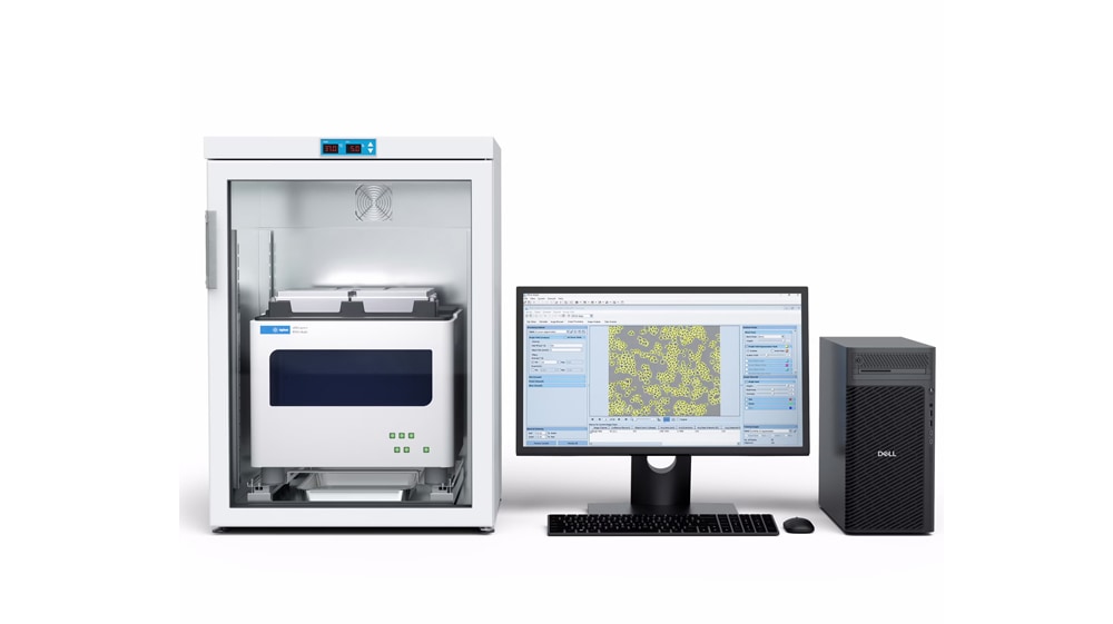
BioTek Gen5 Software for Imaging & Microscopy
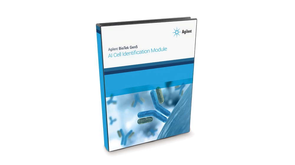
The Agilent BioTek Gen5 AI cell identification module brings AI-based segmentation into familiar Gen5 imaging workflows for compatible BioTek automated imagers. By reducing conventional threshold setting and iterative parameter tuning, the module helps simplify image analysis and support more consistent cell counting, confluence, and morphology measurements across supported fluorescence, brightfield, phase, color brightfield, and confocal images, enabling researchers to move from image acquisition to quantitative results with less manual effort.
Webinars
A New Outlook on Real-Time Cell Analysis: Combining Impedance with AI-Powered Imaging
Traditional endpoint assays provide only a snapshot of cellular response, while imaging alone may leave important biological context unresolved. In this webinar, discover how the Agilent xCELLigence RTCA eSight platform combines real-time impedance with live-cell imaging from the same well to provide a more complete view of cellular function and behavior.
The session will explore how AI-powered, label-free image analysis transforms brightfield imaging into a scalable quantitative workflow, automatically segmenting cells and tracking cell count, confluence, proliferation, and treatment response over time without dyes, parameter tuning, or user-dependent thresholds. Attendees will see how AI-derived imaging measurements complement real-time impedance data from the same cells, providing independent optical and functional perspectives on when cellular responses occur and how they progress. The webinar will also examine how live-cell imaging and kinetic analysis support complex 3D spheroid and organoid studies.
Attendees will learn how to combine imaging and impedance data to support reproducible, scalable workflows in drug discovery and translational research.
Learning objectives
- Understand the complementary functional and visual information provided by real-time impedance and live-cell imaging
- See how AI-powered, label-free image analysis enables automated, reproducible quantification of cellular changes over time
- Explore applications of live-cell imaging and kinetic analysis across 2D and 3D cell models
Speaker

Ryan Raver, PhD | Senior Product Manager, xCELLigence RTCA, Cell and Biomolecular Analysis Division (CBD) | Agilent Technologies.
Upcoming Conferences
SITC 2026
Cardiovascular Research
SFN 2026
November 14‒18, 2026 | Washington, DC
Cell & Gene Therapy Analytical Development Summit
December 1‒3, 2026 | Boston, MA
Cell Bio 2026
December 12–15, 2026 | San Diego, CA
Antibody Engineering & Therapeutics 2026
December 13‒16, 2026 | San Diego, CA
Promotions
Talk to your Agilent representative to find out about promotions in your area.