Choosing the Right Imaging Microplate
September 2025
Microplates, otherwise known as multiwell plates, or microtiter plates, are an essential and broadly utilized sample format for diverse research applications. As research procedures, instrumentation, and throughput demands have changed over time, microplates have adapted, including the development of specialized formats to support automated imaging applications. With the increasing number of imaging microplates now available, it can be challenging to identify which offering is most appropriate for a given use case.
Common questions to consider include the following:
- Can the experiment be performed using label-free imaging, or will fluorescent labels need to be incorporated? What type of microplate is appropriate for each?
- What magnification is necessary to view the results of the image-based test procedure? Will the bottom thickness of the plate well be suitable for use with the desired microscope objective?
- What microplate is appropriate for confocal imaging?
- If three-dimensional (3D) models will be incorporated, what microplate yields the best images with these models?
In this edition of TekTalk, we will explore solutions for the questions listed above.
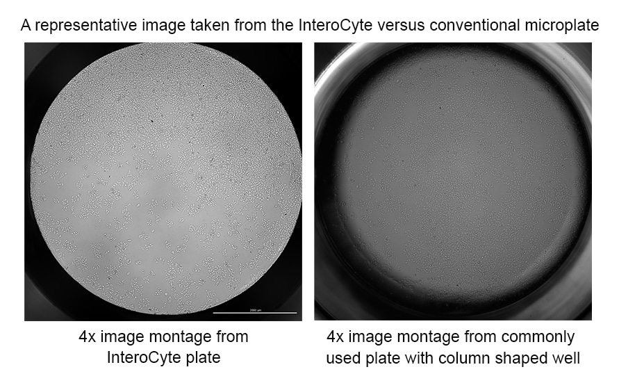
Many image-based workflows that are commonly performed across a wide range of research areas include examining how target cells proliferate. A common desire is the ability to properly image and analyze all cells in the well to accurately assess the efficacy or potential toxicity a test molecule has on the target cells. Microplates commonly used in research today incorporate a column-shaped well, which complicates this goal, due to the dark shadow seen around the well edge from the liquid meniscus. The effect makes accurate cell count or confluence measurements nearly impossible. Agilent InteroCyte shadow-free imaging microplates was developed to alleviate this problem. The unique well design eliminates the shadow caused by the meniscus from the imaged area which contains the cells.
When incorporating high magnification imaging (20–60×), microplates which incorporate a thicker well bottom (500–1000 µm) may not yield the image clarity required to see smaller stained areas of interest, such as mitochondria, muscle fibers, biomarkers, etc. The same is true when working with 3D models, such as spheroids or organoids. The thickness of the bottom can interfere with accurately visualizing each individual slice of the 3D structure. A microplate that includes a film bottom similar to the thickness of a microscope slide coverslip (170–190 µm), such as Agilent’s 204626-100 microplate, can aid in providing the resolution required for these imaging workflows.
Finally, the ability to perform image-based high content screening, in a high throughput manner, remains an important part of the drug discovery process. With compound and large molecule libraries numbering in the tens to hundreds of thousands, higher density plate formats are required to screen each molecule, while reducing the number of plates to process. 384-well microplates, which still incorporate optically clear film bottoms of coverslip thickness, such as Agilent’s 204628-100 microplate, provide an ideal solution to perform high-throughput 2D or 3D cell-based assays.
Featured Application Notes and Technical Overview
Whole-Well Cell Proliferation Imaging with the Agilent InteroCyte Shadow-Free Imaging Microplate
This technical overview explains how the incorporation of a microplate, which enables equal illumination of cells across an entire well when using transmitted light imaging, provides high quality images and consistent data analysis.
Automated Assay for Measuring CAR T Cell Killing in 3D Cancer Models
In this application note, discover how Agilent imaging plates were used to perform hydrogel-embedded 3D T cell killing assays with the Agilent BioTek Cytation C10 confocal imager reader.
Image-Based Cell Cytotoxicity Screening Using Agilent 384-Well Microplates
This application note describes the use of the Agilent 384-well clear-bottom, black-sided cell culture and imaging plate for cytotoxicity screening assays. Low background fluorescence allows the use of common cytotoxicity reagents, while the tissue culture-treated substrate provides an excellent surface for cell growth and supports long-term, cell-based assays.

Automated High-Throughput Imaging and Analysis of Cell Migration
In this application note, learn about the advantages of using the Agilent BioTek Gen5 object tracking module in concert with the Agilent BioTek Lionheart FX automated microscope, and Agilent 96-well imaging plates, for conducting high throughput 2D cell migration studies.
Tek Tips
- When performing kinetic cell proliferation assays, to avoid overpopulation of cells within the well, optimize initial cell seeding concentrations to account for the specific growth rate of the target cell model.
- Before imaging, ensure that the correct z-height is established in the microscope/imaging system software to properly view the cells in focus, for the specific plate type being used. This is the distance in the z-axis between the bottom of the plate skirt and the interior of the well bottom, or bottom elevation.
- If a multiday live-cell imaging assay procedure is being carried out, be sure to include media exchanges to ensure that proper nutrients and growth conditions are being supplied to the cells at all times.
- If high magnification, or confocal imaging, will be incorporated into the experimental procedure, to capture the most in-focus images, it is recommended to use a microplate with an optically clear film bottom of coverslip (170–190 µm) thickness.
- When performing full well label-free imaging (brightfield, high-contrast brightfield, phase contrast), it is recommended to use a plate which can provide consistent illumination across all areas of the well, to achieve accurate cell count or confluence determinations.
Product Spotlights
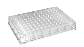
Agilent InteroCyte Shadow-Free Imaging Microplate
The Agilent InteroCyte shadow-free imaging microplate incorporates a unique well design which eliminates liquid meniscus shadows seen around the edge of a microplate well. This feature provides accurate cell count and confluence measurements when performing full well label-free imaging, perfect for uneven or low cell densities. The versatility of the plate also enables its use for non-full well label-free, as well as fluorescence imaging applications.

BioTek Cytation C10 Confocal Imaging Reader
The Agilent BioTek Cytation C10 is a versatile confocal imaging reader designed for high-resolution imaging of complex biological systems. With its spinning disk confocal technology and water immersion objectives, the Cytation C10 delivers exceptional clarity for many different sample types. With its unique flexibility, the Cytation C10 is compatible with all standard SBS/ANSI microplates from 6- to 1536-well, including the new Agilent InteroCyte shadow-free imaging microplate. This flexibility allows you to seamlessly integrate the system into your existing lab workflows.
Additional Resources
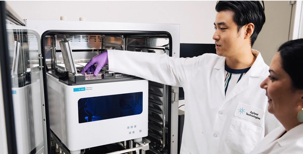
xCELLigence RTCA eSight
The Agilent xCELLigence RTCA eSight is an advanced real-time cell analysis system that enables comprehensive insight into a cell's health, behavior, function, and biological processes using live, simultaneous, and real-time biosensor-based and image-based measurements. The xCELLigence RTCA eSight system combines label-free impedance technology with flexible live-cell brightfield imaging and three fluorescent channels spanning a multitude of microplate forms (6-, 12-, 24-, 48-, 96-, or 384- well, including Agilent InteroCyte shadow-free imaging microplate).
Agilent Microplate and Reservoir Introduction Video
Gain a more accurate picture of how Agilent’s extensive portfolio of reservoirs, storage plates, and microplates provide solutions to setup and perform a wide variety of biochemical and cell-based assay procedures.
Scientific Conference
Join us at CAR-TCR Summit in Boston (Sept 23 – 26)