Organoids and 3D Models
March 2026
Over the past decade, there has been a rapid expansion in the use of three-dimensional (3D) cell culture models, including organoids, across medical research, precision medicine, disease modeling, and drug discovery. These advanced cell culture systems are becoming valuable tools in preclinical research given their capacity to more accurately replicate the structural and functional characteristics of real human tissues, overcoming many of the limitations of traditional two-dimensional (2D) cell culture models.
Importantly, the U.S. Food and Drug Administration (FDA) roadmap to reduce reliance on animal testing emphasizes the need for validated new approach methodologies (NAMs). Organoids and other 3D models are a key factor of this strategy, as they offer physiological insights that can enhance the translational value of research while reducing the need for animal testing.
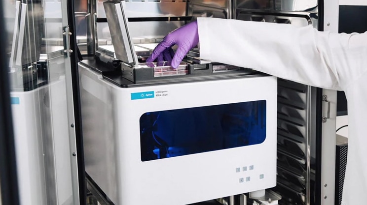
In alignment with these FDA priorities, Agilent has developed a broad range of assay workflows, integrating powerful and unique analysis platforms with organoids and other 3D models in mind. In this issue of TekTalk, we highlight several of these innovations, including the Agilent Seahorse XF Flex analyzer and Agilent Seahorse XF Flex organoid microplates which‒together‒enable comprehensive metabolic profiling of organoids. We also feature the Agilent xCELLigence RTCA eSight, which combines label free impedance measurements with live-cell imaging to support real time monitoring of organoid health, morphology, and behavior.
To complement these functional analyses, we highlight imaging strategies tailored for organoids and 3D models, which pose challenges due to greater thickness and optical complexity. The Agilent BioTek Cytation systems with Gen5 software meet these challenges by enabling straightforward low magnification screening and characterization of 3D samples alongside high resolution imaging that delivers clearer optical sectioning and detailed insights into cell model function and treatment response.
Additionally, plate reader–based biochemical assays, including ELISAs, provide robust and scalable orthogonal characterization of 3D systems. Agilent BioTek plate readers offer industry leading sensitivity and flexible detection modes, supporting high performance biochemical and immunoassay workflows that seamlessly complement imaging and real time analyses.
Featured Applications
Metabolic Profiling Cancer Organoids Cultured in the Agilent Seahorse XF Flex Organoid Microplate
This application note describes the use of the Agilent Seahorse XF Flex organoid workflow for the metabolic profiling of cancer organoids using the Agilent Seahorse XF Flex analyzer and Agilent Seahorse XF Flex organoid microplate. The workflow was evaluated by examining the impact of metformin on mitochondrial function in cancer cell-derived organoids. This approach enables screening and characterization of drugs targeting mitochondrial function in 3D culture models such as cancer organoids.
High-Throughput Methods to Quantitatively Evaluate TGF-β Signaling in Epithelial-to Mesenchymal Transition
We detail a high-throughput method for evaluating TGF-β/SMAD signaling in an A549 3D tumor model. Using confocal imaging on the Agilent BioTek Cytation C10, the study simultaneously quantifies biochemical markers, nuclear translocation of p-SMAD2/3 and SMAD4, and phenotypic EMT responses. A direct correlation between the magnitude of nuclear SMAD4 signal and the extent of cell migration from the spheroid core can be seen. This workflow shows how to effectively link intracellular signaling to morphological changes within a complex 3D multicellular model.
Tek Tips
Organoids and 3D Cell Culture Models in Cellular Analysis
Different 3D systems such as spheroids, organoids, and scaffold-based cultures capture distinct aspects of tissue physiology, so defining the biological question and analytical endpoint is essential before selecting a model. Key considerations include:
- Ensure the matrix and media support the intended signaling environment: Growth factor levels, ECM composition, and mechanical stiffness strongly influence differentiation, proliferation, and phenotype.
- Account for nutrient and oxygen gradients:—Intrinsic gradients shape metabolism, drug penetration, and cell behavior; data interpretation should reflect these nonuniform microenvironments.
- Standardize culture geometry and size: Consistent spheroid or organoid dimensions significantly improve reproducibility across imaging, functional assays, and biochemical readouts.
- Confirm analysis method compatibility: Imaging, sequencing, and functional assays may require adjustments based on organoid size, morphology, media components, and ECM type.
Key Experimental Considerations for Cell-Based Assays Using ECM Scaffolds
Many organoid systems use ECM gels or coatings. For reliable, reproducible analysis, focus on the following high-impact practices:
- Select ECM materials aligned with the biology: Stiffness, protein composition, and crosslinking must match the tissue or functional endpoint being modeled.
- Use defined ECM protein concentrations: Gel stiffness depends on total protein content, so follow provider concentration guidelines rather than relying on dilution ratios.
- Prevent bubbles and structural defects: Quick spin down and slow dispensing reduce defects that compromise imaging, diffusion, and uniform culture architecture.
- Verify assay compatibility early: Check for autofluorescence, reagent binding, and extraction efficiency using matrix only controls; use phenol-red-free media when necessary.
Key Experimental Considerations for Staining 3D Cell Models and Organoids
Staining 3D cultures requires strategies that address limited diffusion, dense ECM, and fragile sample structure. The following high impact practices help ensure consistent, high quality labeling.
- Optimize stain penetration: Use mild permeabilization (e.g., low percentage Triton X 100 or saponin) and extend staining to multi hour or overnight incubations. Rock or rotate samples to reduce diffusion gradients and verify depth uniformity with Z stacks.
- Select stains compatible with 3D biology and ECM: Choose low molecular weight or membrane permeant dyes to improve penetration and reduce matrix binding. Validate functional probes‒such as mitochondrial, reactive oxygen species (ROS), or apoptosis indicators‒directly in 3D, where gradients can influence probe behavior. Favor far red fluorophores to minimize scattering.
- Use staining tools designed for 3D workflows: Agilent eLive dyes and the xCELLigence eSight imaging platform provide robust, multiplexed live cell labeling optimized for the diffusion and optical challenges of spheroids and organoids.
- Protect structure and signal during preparation and imaging: Handle samples gently using wide bore tips and minimal pipetting; stain in plate whenever possible. For endpoint assays, fix early and use mild clearing solutions validated on a small test set. Reduce photobleaching with anti fade reagents and low excitation power.
Product Spotlights
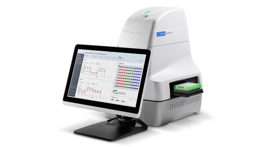
Seahorse XF Flex with XF Flex Organoid Microplates
The Agilent Seahorse XF Flex enables the real-time measurement of cellular bioenergetics across diverse 3D study models, including organoids and tissue materials. In addition, the Agilent Seahorse XF Flex organoid microplate is optimized for matrix-embedded organoid culture, allowing long-term growth, high-resolution imaging, and comprehensive metabolic profiling within the same well. Together, they allow high resolution, functional metabolic profiling of organoids and other 3D models, enhancing translational relevance in disease research and drug screening.
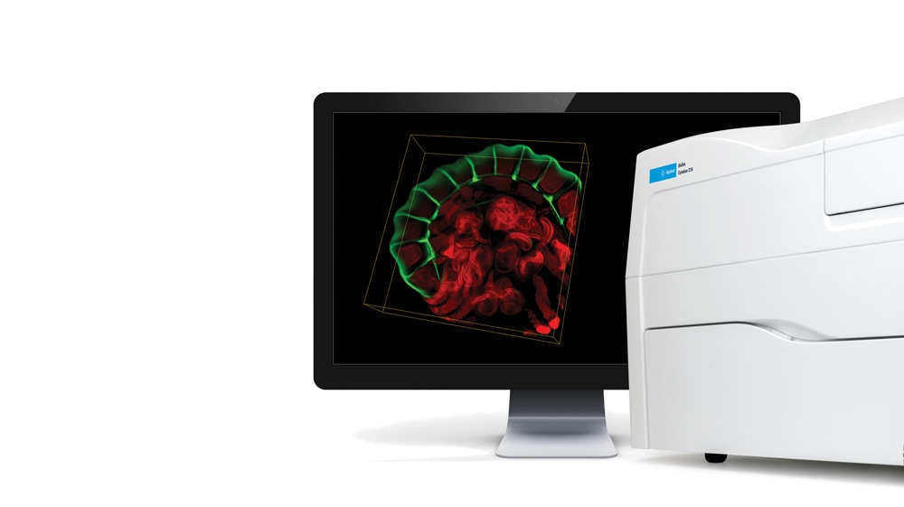
BioTek Cytation C10 Confocal Imaging Reader
Master complex 3D biology with the advanced optical sectioning capabilities of the Cytation C10 confocal imaging reader. The combination of automated confocal microscopy with multimode detection allows this system to capture deep Z-stacks to quantify nuclear translocation within thick 3D models while simultaneously measuring biochemical outputs. Experience the versatility needed to correlate subtle intracellular behavior with phenotypic outcomes in a single high-throughput workflow.
xCELLigence Real-Time Cell Analysis Systems
Agilent xCELLigence Real-Time Cell Analysis (RTCA) systems use noninvasive biosensor technology to provide continuous, quantitative monitoring of cell health and behavior—ideal for environmental and water quality applications. This platform enables real-time assessment of processes such as chemical- and contaminant-induced cytotoxicity, with minimal hands-on effort. By delivering highly sensitive kinetic data, xCELLigence RTCA surpasses traditional endpoint assays, offering deeper insights into dynamic cellular responses to pollutants and pathogens.
Featured Video
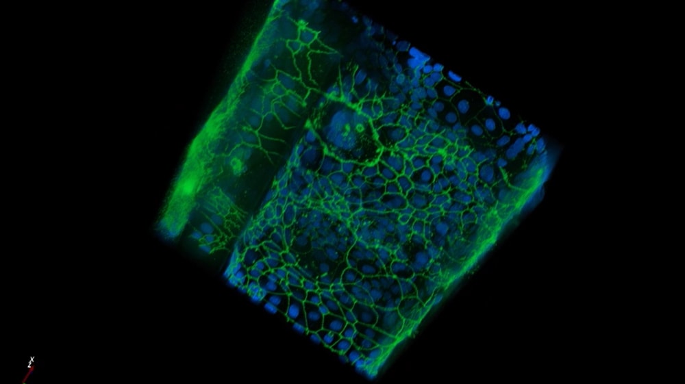
3D Rendering of an Intestinal Organoid
A Z-stack and 3D rendering of an intestinal organoid model imaged with the spinning disk confocal optics of a Cytation C10. Caco-2 cells seeded and allowed to form a tube in the Mimetas OrganoPlate.
Webinar
Confidently generate human-relevant metabolic data from 3D organoid models using the Agilent Seahorse XF Flex organoid workflow
In alignment with FDA’s priority focused on new approach methodologies (NAMs), Agilent developed an assay workflow to interrogate energy metabolism and assess mitochondrial function in organoid cultures using the XF Flex organoid microplate and the XF Flex analyzer. This workflow empowers physiologically relevant discoveries in a wide range of biomedical research areas.
In this webinar, you will learn:
- How to establish organoid cultures in the XF Flex organoid microplate and perform Seahorse XF assays.
- Techniques for optimizing assay performance with different types of organoid cultures and data normalization options
- Examples demonstrating the applicability of the XF Flex organoid workflow in cancer research and drug discovery as well as in drug safety studies.
Additional Resources
Application & Industries
Application Notes
- Confocal Imaging and Analysis of Spheroids for Determination of Dose Response during Drug Treatment
- A Superior System for Real-Time Metabolic Analysis with Brain Tissue and Other 3D Models
- Making Barrier Integrity Assays Easier: Automated Imaging of Intestinal Barrier Function on a High-Throughput Organ-on-a-Chip Platform
- Three-Dimensional Multispheroid Assays Using the Agilent xCELLigence RTCA eSight
- Monitoring Drug-Mediated 3D Tumor Spheroid Shrinkage in Real Time Using the Agilent xCELLigence RTCA eSight
Technical Overviews
- Measuring Mitochondrial Function of Matrix-Embedded Organoids Using the Agilent Seahorse XF Flex Analyzer
- Immune Cell Killing of 3D Tumor Spheroids Using the Agilent xCELLigence RTCA eSight
- Real-Time Monitoring and Quantifying 3D Tumor Spheroid Invasion Using the Agilent xCELLigence RTCA eSight System
Poster
Upcoming Conference
SOT 65th Annual Meeting and ToxExpo
March 22-25, 2026 | San Diego, CA
April 17-22, 2026 | San Diego, CA
xCELLigence Cancer & Immunotherapy Symposium
April 18, 2026 | San Diego, CA