Gen5 Object Tracking
January 2023
The concerted movement of cells within their environment plays a fundamental role in nearly all eukaryotic organisms. Higher eukaryotes rely on regulated cell migration throughout development and for immunologic processes, while aberrant cell migration is a defining feature of metastatic cancers. The process of cellular migration towards external stimuli (such as chemotaxis) is called directional cell migration. Equally as informative is random cell migration, in which cell trajectories are measured when a chemoattractant is diffuse instead of emanating from a source direction. Whether studying directional or random migration, it is important to measure the motile capacity of cells accurately and reliably.
The Agilent BioTek Gen5 Object Tracking Module provides an automated method for characterizing 2D cell migration in high-content formats, including 96-well microplates and chemotaxis slides. Combined with Agilent BioTek automated imaging systems, this capability delivers an integrated solution for automating cell migration studies, increasing assay robustness and throughput. Below we discuss a few aspects to consider when setting up both random and direct cell migration assays.
Featured Applications
Automated High-Throughput Imaging and Analysis of Cell Migration
The ability of cells to migrate is a fundamental biological process in development and immunologic responses. It is also the defining feature of metastatic cancers. Therefore, it is important to measure the motile capacity of cells accurately and reliably. This application note introduces the Agilent BioTek Gen5 object tracking module, which automates analysis of 2D cell migration in high-content formats, including 96-well microplates.
Automated Imaging and Analysis of 2D Chemotaxis
The ability of cells to migrate is a fundamental biological process in development and immunological responses. It is also the defining feature of metastatic cancers. The process of cellular motility towards external cues is collectively directional cell migration, for which migration towards a soluble chemical stimulant is referred to as chemotaxis. Measuring chemotaxis requires the ability to track cellular movement accurately and reliably.
Product Spotlight
Agilent BioTek Gen5 Software for Imaging & Microscopy
BioTek Gen5 Software for imaging and microscopy offers automated image capture, processing, and analysis for a broad range of samples, from whole organism imaging to high magnification subcellular imaging. Raw data and images are transformed into meaningful results with powerful built-in capture, process, and analysis tools. The automated process, known as Augmented Microscopy, enables simple and straightforward imaging and analysis workflows.
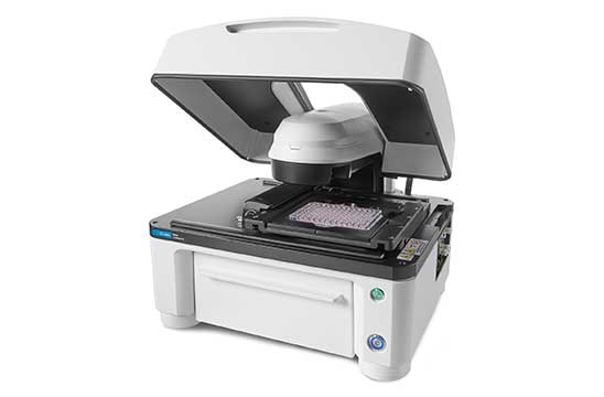
Agilent BioTek Lionheart FX Automated Microscope
Lionheart FX Automated Microscope offers up to 100x magnification with brightfield, color brightfield, phase contrast and fluorescence channels for fixed and live cell applications. Optional incubation to 40°C, CO2/O2 control, and a humidity chamber optimize live cell assays. Gen5 software makes cell image capture, processing, analysis, and publication-ready data and images quick and effortless.
Videos
Agilent BioTek Gen5 Object Tracking Module
Tek Tips
General tips for conducting migration assays
Conduct migration assays using a culture vessel coated with an appropriate extracellular matrix
2D and 3D cell migration is more efficient in the presence of an extracellular matrix (ECM) than on bare glass or even TC-treated plastics. This is because the most common form of motility in eukaryotic cells is amoeboid movement, which depends on stable adhesion between a cell and its surrounding environment. Cells accomplish this task through integrins, which are heterodimeric transmembrane receptors that directly bind to components of the ECM (REF#1). Integrin receptors are composed of an α and β subunit and the specific α/β isoform combination confers affinity for one ECM protein over another, which in turn influences how well a particular cell type can migrate through its environment. For example, the α5β1 integrin preferentially binds to fibronectin, whereas α1β1 binds collagen, and α3β1 binds laminin (REF#2). Therefore, choosing the appropriate ECM to use for in vitro migration studies largely depends on the integrin expression profile of your cell model. However, if defining a cell model’s integrin expression profile is not practical, an initial pilot study is recommended where cells are seeded on different ECMs, and their adhesive and motile potential is evaluated. Equally as important is to determine the ECM coating concentration for either one that maximizes migration potential, or one that best reflects in vivo concentrations of a cell’s local microenvironment.
Optimize cell seeding density
One can imagine that a cell can only reach its maximal motility rates when it is unobstructed by another object in its path. Additionally, it can be argued that once a cell comes up and interacts with another cell, its trajectory has inherently been altered and subsequent kinetic data is invalid. Therefore, choosing a seeding density should be influenced by the following: 1) the lag time between seeding and experiment execution, 2) the motility rates of the cells, and 3) the duration of the kinetic run.
Chemotaxis assay optimization
Determine the chemoattractant response range
Before undertaking a chemotaxis assay, it is important to establish the responsive chemoattractant concentration range for your particular cell model, which would then dictate the gradient range in the chemotaxis chamber. This can be accomplished with a pilot random migration assay in a 96-well microplate, where serum-starved cells are treated with a dilution series of chemoattractant, and an EC50 is established. This EC50 can then help decide the chemoattractant concentration that should be use in the source chamber of a chemotaxis assay.
Verify that a chemoattractant gradient has been established
Interpreting chemotaxis migration data relies on a successfully established chemoattractant gradient in a chemotaxis chamber, and fluorescence microscopy offers the ability to verify the presence of a gradient. The most direct and ideal approach to accomplish this is to use a fluorescently conjugated chemoattractant. If a conjugated form of a chemoattractant is not available, an alternative approach is to use a secondary molecule of comparable molecular weight which is conjugated to a fluorophore. Tetramethyl rhodamine (TRITC)-conjugated Dextran can be used for this purpose and is available in a variety of molecular weights (REF#3). Imaging this fluorescently visible gradient in a kinetic run will also provide insight to the stability of a gradient over the course of the chemotaxis duration. Lastly, once the gradient is verified, it is possible to then correlate the region within gradient where a response is most likely to occur, based on the EC50 previously established in a pilot dilution series migration assay.
References
- Zaidel-Bar, R. Job-splitting among integrins. Nat Cell Biol 15, 575–577 (2013). https://doi.org/10.1038/ncb2770
- Humphries, et al. Integrin ligands at a glance. J Cell Sci 1 October 2006; 119 (19): 3901–3903. https://doi.org/10.1242/jcs.03098
- Wu et al. Arp2/3 Is Critical for Lamellipodia and Response to Extracellular Matrix Cues but Is Dispensable for Chemotaxis. Cell 148(5), 973-987 (2012). https://doi.org/10.1016/j.cell.2011.12.034.
Resources
Webinar
Agilent BioTek Gen5 Object Tracking Module: Applications in 2D cell migration
Wednesday, January 25, 2023 | 12PM EDT
Presenter: Ernest Heimsath, Ph.D.; Application Development Scientist, Agilent Cell Analysis
The ability of cells to migrate is fundamental for many biological processes and is also the defining feature of metastatic cancers. When quantitatively analyzing cell migration behavior, it is important that their movement is recorded accurately and reliably. In addition, there are a few experimental aspects to consider when planning a live-cell imaging session for the purposes of tracking cell movement. The Agilent BioTek Gen5 object tracking module automates analysis of 2D cell migration, whether it is random migration, or directed migration.
In this training webinar, we’ll cover:
- Tips and tricks for setting up and conducting cell migration experiments
- Best practices to ensure successful implementation of the Gen5 object tracking module
- Explore a number of cell migration applications and analysis
Promotions
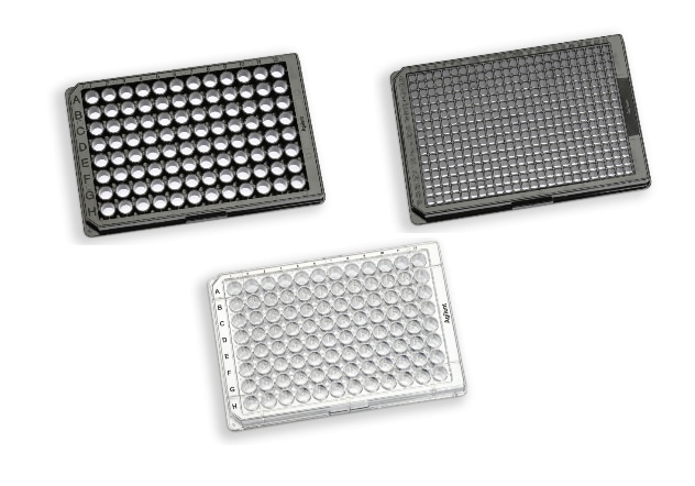
Complete Your Cell Based Assay Workflow with Agilent Microplates
Sourcing your microplates shouldn't be an added task in preparing your cell-based workflows. Agilent now offers cell culture and imaging microplates so you can complete your workflows seamlessly.
Expect the quality you need with TC surface treatment enhancing the cell attachment while the microplate lid which accompanies every plate maintains a sterile growth environment with a low risk of cross contamination.