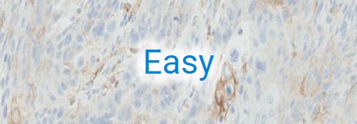
Atlas of Stains: PD-L1 IHC 22C3 pharmDx

The Atlas of Stains: PD-L1 IHC 22C3 pharmDx is a digital repository of NSCLC tissue samples stained with PD-L1 IHC 22C3 pharmDx including:
- Positive cases that span the full range of PD-L1 expression
- Negative cases that may demonstrate intrinsic controls (tumor associated macrophages and immune cell staining)
- Full specimen staining with H&E, primary antibody, and Negative Control Reagent (NCR) for each patient
The viewer interface for the Atlas of Stains: PD-L1 IHC 22C3 pharmDx features:
- High-definition, zoomable scans with full-screen and quadrant-view functionality for detailed PD-L1 stain analysis
- Expert annotations describing areas of interest plus the ability to add your own annotations
- Tumor Proportion Score (TPS) for each stain, to verify your own assessment


For in vitro diagnostic use
PD-L1 IHC 22C3 pharmDx is a qualitative immunohistochemical assay using Monoclonal Mouse Anti-PD-L1, Clone 22C3 intended for use in the detection of PD-L1 protein in formalin-fixed, paraffin-embedded (FFPE) non-small cell lung cancer (NSCLC) tissue using EnVision FLEX visualization system on Autostainer Link 48.
PD-L1 protein expression is determined by using Tumor Proportion Score (TPS), which is the percentage of viable tumor cells showing partial or complete membrane staining. The specimen should be considered PD-L1 positive if TPS ≥ 50% of the viable tumor cells exhibit membrane staining at any intensity.
PD-L1 IHC 22C3 pharmDx is indicated as an aid in identifying NSCLC patients for treatment with KEYTRUDA® (pembrolizumab).