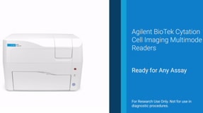
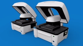
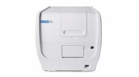
Cell Imaging & Microscopy for Automated Image Capture and Analysis
Agilent BioTek automated cell imagers and microscopes bring your science to life, capturing spectacular images, z-stacks, montages and time lapse sequence with ease. With up to 100x magnification, plus brightfield, color brightfield, phase contrast and fluorescence channels, these instruments support a wide range of microscopy workflows including live cell kinetics. The compact, modular systems automate image capture, process, and analysis workflows to meet most laboratory budgets. Capture modes in the product range include fluorescence, brightfield, high contrast brightfield, phase contrast, and color brightfield. Agilent BioTek Gen5 software controls the instrumentation and provides intuitive programming and analysis.
Discover Agilent's automated imaging-based method for robust analysis of CAR T cell cytotoxicity, enhancing cancer treatment efficacy and research efficiency.
Rigorously validated antibody reagents from Cell Signaling Technology, combined with the multifunctional capabilities of the Agilent BioTek Cytation C10 confocal imaging reader, enable robust, quantitative analysis of TGF-β and BMP pathway activation at different biological scales.
This application note describes whole-organism imaging of spinal motor neurons and musculature using the Agilent BioTek Cytation C10 confocal imaging reader.
Join our webinar to learn how Boehringer Ingelheim uses AVIA AI and Cytation imaging to automate viral infectivity assays and speed up vaccine development.
Hepatocarcinoma is one of the most common cancer types in the U.S. with limited treatment options and poor survival rate. Recently, cancer bioenergetics emerged as a promising source of targets to develop novel anticancer agents.