Image Gallery
Agilent BioTek imaging products are designed for a wide range of applications. Our customers are using the Lionheart Automated Microscopes, and Cytation Cell Imaging Multi-Mode Readers for their own unique research and in the process acquire beautiful, publication quality images and movies. Take a look at images captured with Agilent BioTek instruments.

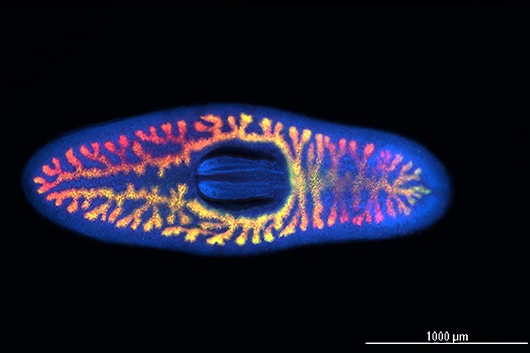
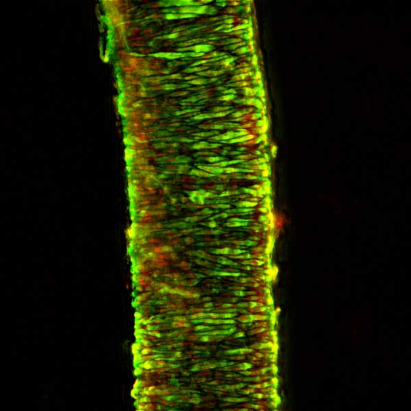
 and AcTub (green) to specifically stain the ciliated cells with DAPI (blue) labelling nuclei. Captured with the Agilent BioTek Cytation 5 cell imaging multimode reader")
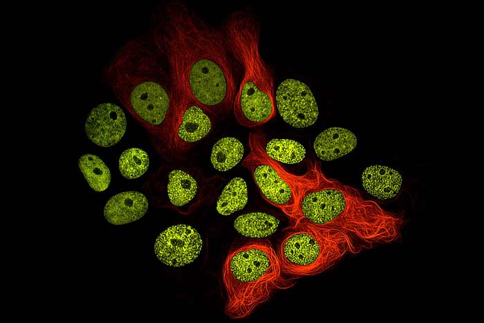
 that has been fixed and stained for Nuclei (Blue; Hoechst 34580), Microtubules (Green; anti-alphatubulin), Mitochondria (Red; anti-Tom20), and F-actin (Gray; phalloidin). Captured with the Agilent BioTek Cytation C10 confocal imaging reader.")

, MAP2 (white) and counterstained with DAPI (blue). Captured with the Agilent BioTek Cytation C10 confocal imaging reader.")

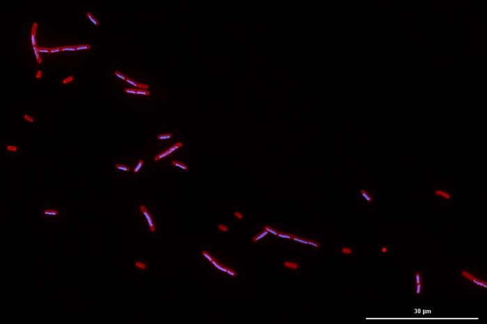
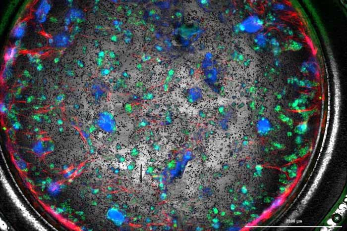
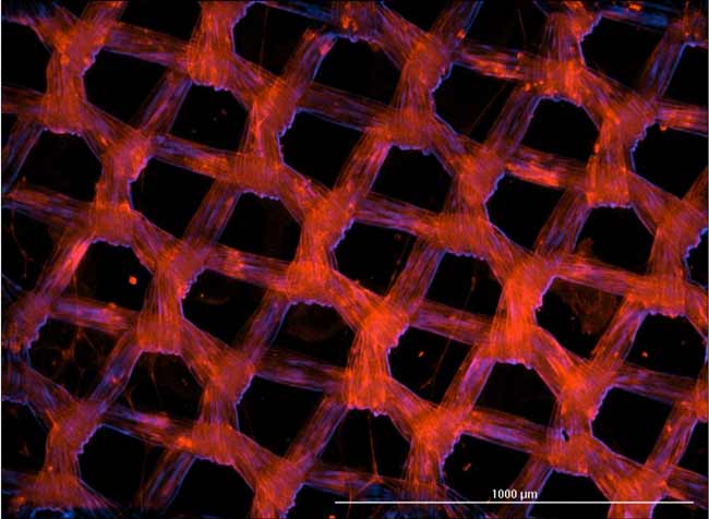
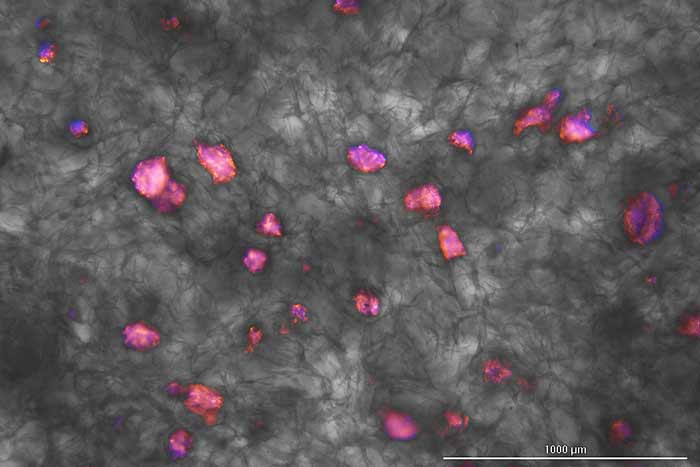
, extracellular polymeric substance (EPS) proteins (red), and EPS polysaccharides (blue). Image shown is a single slice of an image captured on day 8 of development, at 60x confocal with Cytation C10 confocal imaging reader")

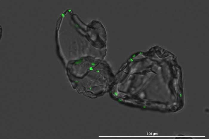
, CF633-conjugated goat anti-rabbit). Captured at 60x water immersion with Cytation C10 confocal imaging reader.")
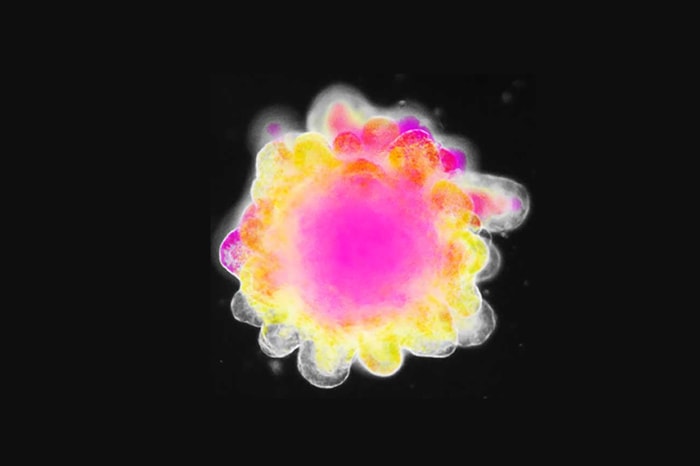
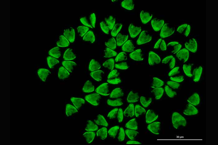
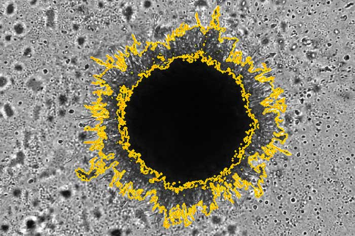
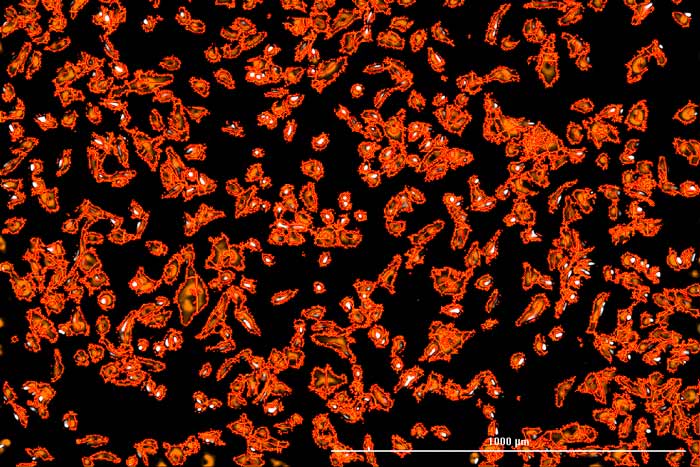
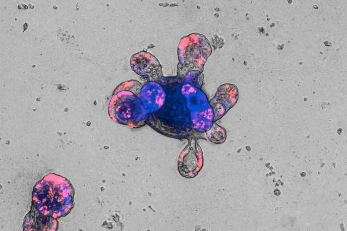
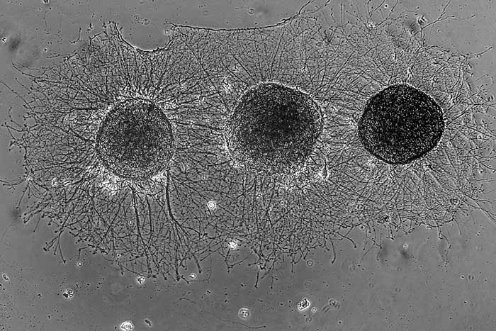
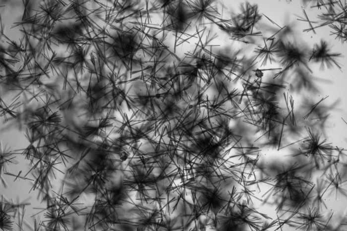
, at 10x brightfield, of a Dictydium cancellatum, a myxomycete of the order Liceales. Pictured is the sporotheca, the bulbous container at the head of the organism that produces and contains the spore mass. Captured at 10x brightfield with Cytation 5 cell imaging multimode reader.")

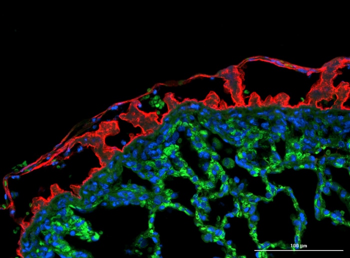
 and counterstained with Alexa Fluor 488-conjugation phalloidin (green) and Hoechst 34580 (blue). Imaged at 60x water immersion. Image captured at 100 µm depth.")
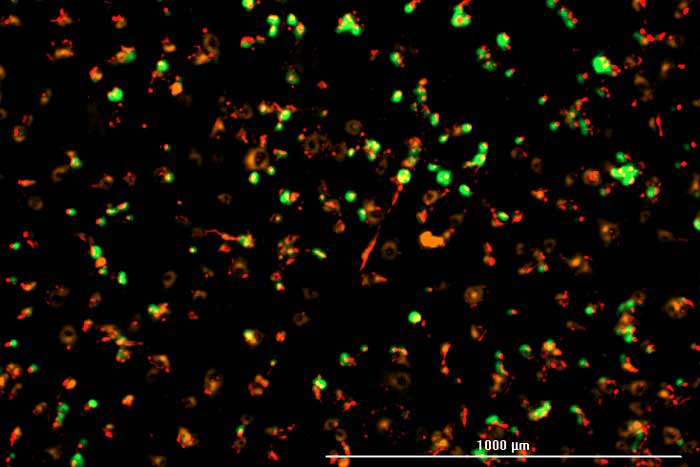
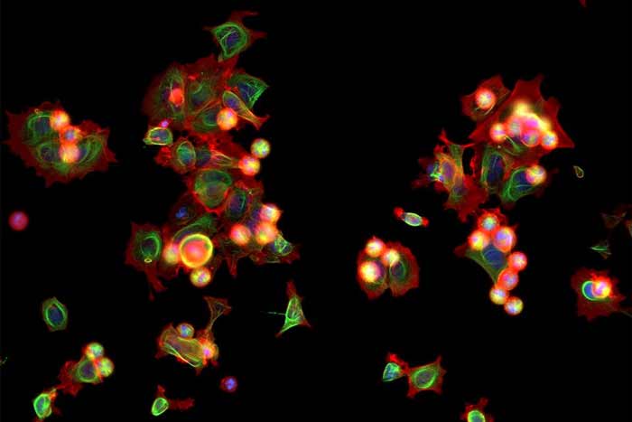
, tubulin (green), nuclei (blue), mitochondria (orange). Widefield 2x2 montage captured at 40x with Agilent BioTek Cytation C10 confocal imaging reader.")


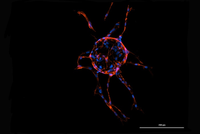
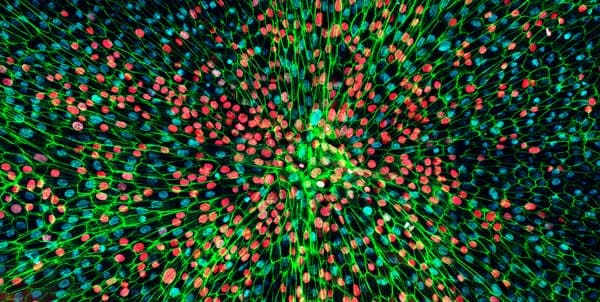
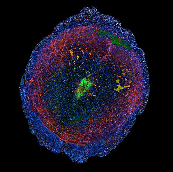
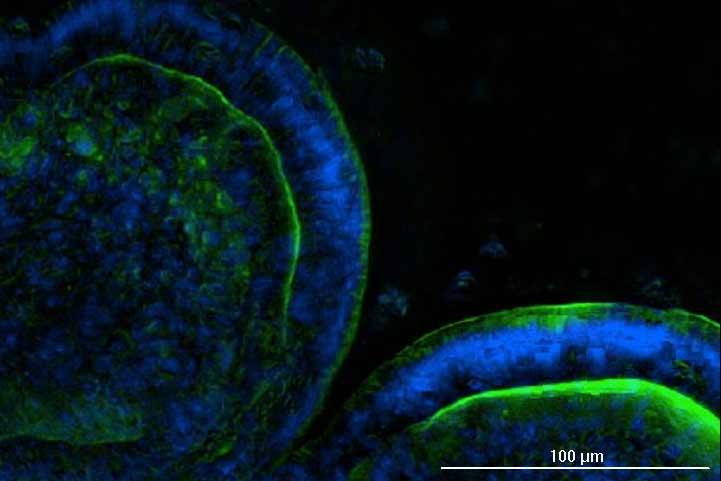
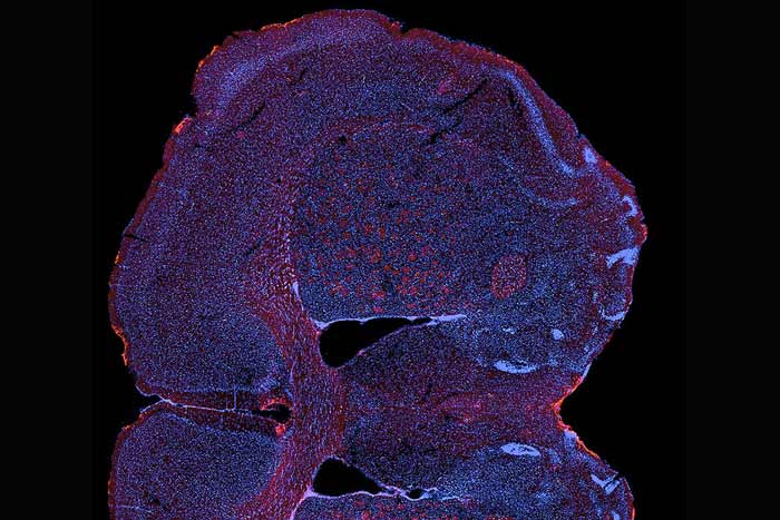
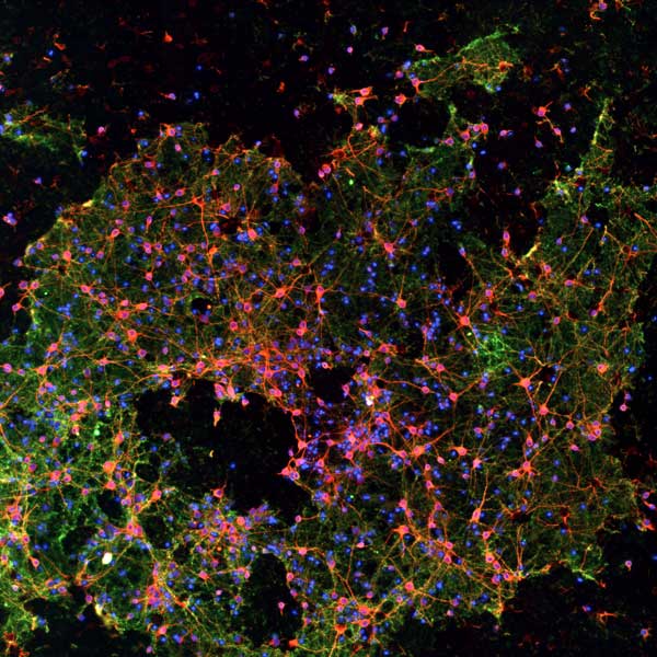
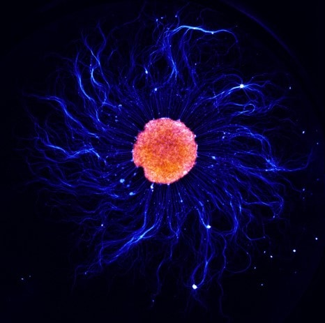
, tyrosine hydroxylase (red) and DAPI (blue). Image captured with Lionheart FX. Summited for the 2021 Imaging contest by Hsueh Fu Wu, University of Georgia")
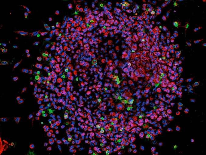
 from eBioscience and nuclei co-staining using DAPI. The image was taken at a 20x magnification. Submitted for the 2021 Imaging contest by Karina Cereceda of Fundación Arturo López Pérez. Image captured with Cytation 5.")
 taken as a different way to look at cells. Image taken at 40X magnification. Image captured with: Cytation 5. Submitted by Alia Mallah, University of Texas at San Antonio.")
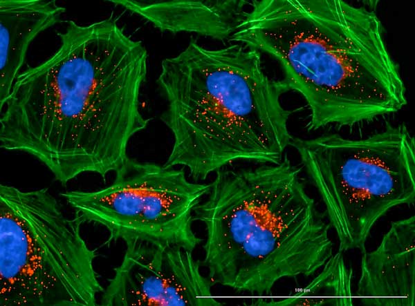
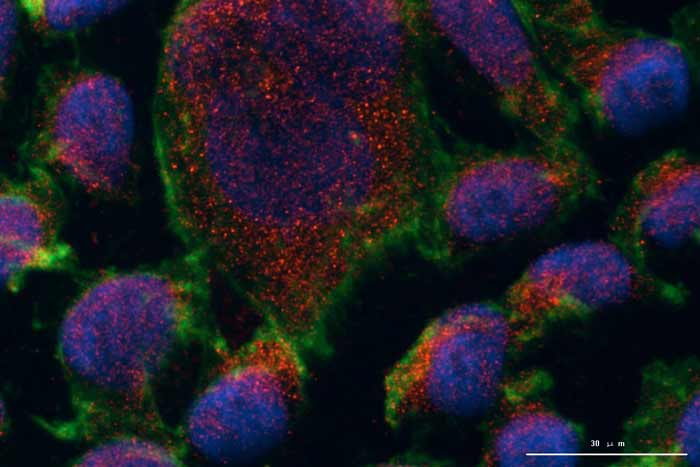
 with the lysosomes (Texas Red channel, anti-LAMP2) which accumulate inside the cells. Image captured with: Lionheart FX by Cristina Andreani, University of Cincinnati")
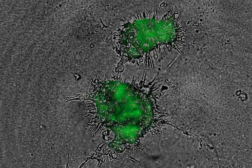
 being targeted and killed by primary human natural killer cell (in red) following treatment with a pro-immunomodulatory agent. Live cell imaging was performed using a Cytation 5 at 10x using two channel fluorescence and brightfield imaging. Image captured with: Cytation 5 by Catherine Mills, Medical University of South Carolina")
, O-GlcNAcylation (red), mitochondria (green), and DAPI. Image was taken at a 10x magnification with automatic image processing. Image captured with: Lionheart FX by Chia-Wei Huang, University of Georgia")
 and fibrin stain (647); moreover, images were taken at 4X. Image captured with: Cytation 5")
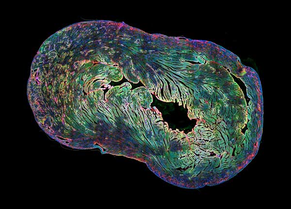
 and H-ATPase (green) in kidney sections of mice. We can observe the cellular conformation of renal tubules and cell nuclei are stained with DAPI (Blue). Image was taken at a 20x magnification. Image captured with: Cytation 1")


, TUJ1 (red), DAPI (blue). Image was taken using a Lionheart FX (manual mode) at a 4X magnification.")

, TUJ1 (green), and DAPI (blue). Image was taken using a Lionheart FX (manual mode) at a 4X magnification.")
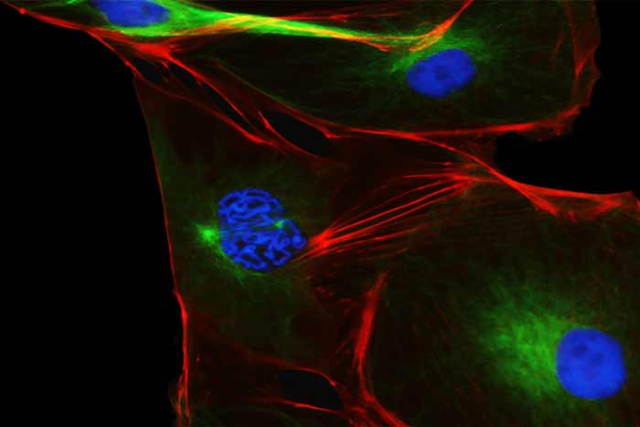
, and different phases of cell cycle can be found in this picture")
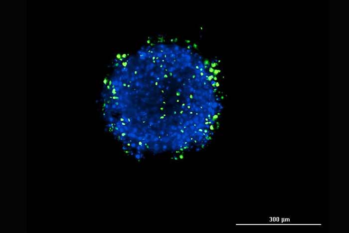
 with z-stacking every 25 um")

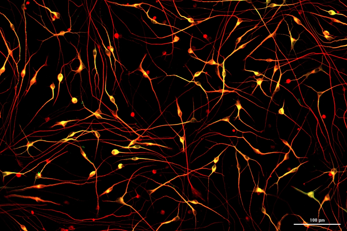
 and neurofilament (Alexa Fluor 647, red) (10X objective).")
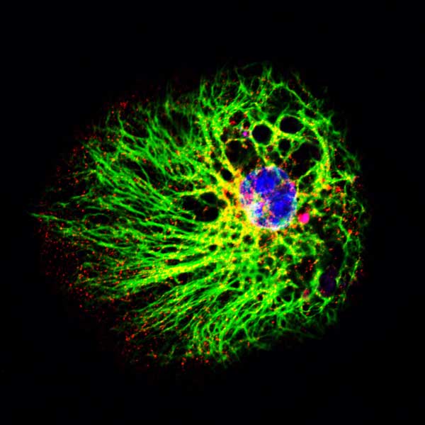
 and lysosome GFP (red). Images were acquired using 60X objective. Image shows Z stack projection of maximum fluorescence intensity.")

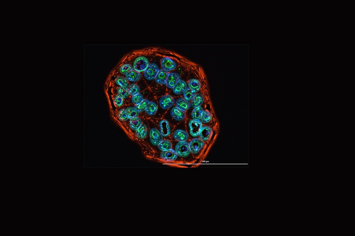
, and Somatostatin (Alexa Fluor® 488), with DAPI stained nuclei, of porcine islet, imaging at 20x.")
 adipocyte cells stained with BODIPY and Hoechst 33342.")
.")
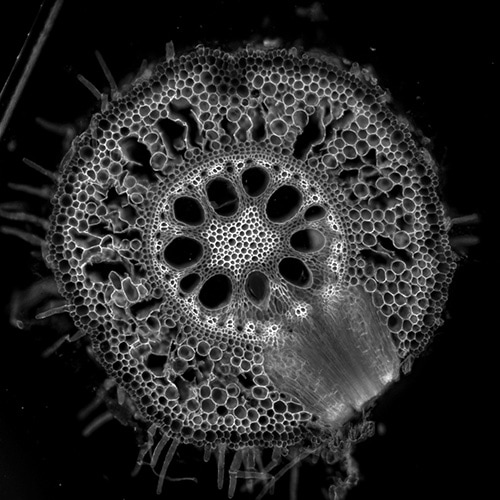
. White/pink color is autofluorescence that indicates vascular cells with enhanced wall thickening. The image was captured at 10x and with brightfield and DAPI, FITC and Rhodamine filters. Captured by Lionheart FX. Submitted for the 2019 Imaging Perspectives contest by Elison Blancaflor, Noble Research Institute LLC.")
, MitoTracker (red) and DAPI (blue). Captured by Cytation 3. Submitted for the 2019 Imaging Perspectives contest by Ibrahim Halil Demirsoy, Luigi Vanvitelli Campania University.")