Tissue And Whole Organism Analysis
July 2025
Assays performed on living cells are increasingly being shifted from two-dimensional single monolayer cell cultures to three-dimensional models such as organoids, tissue, and small organisms. Why? These models better replicate in vivo conditions, thereby enhancing the studies physiological relevance. 3D models can be created artificially, with an increasing number of engineering strategies available, or naturally occurring models such as tissues or whole organisms such as zebrafish embryos and C. elegans are available.
A few key challenges should be considered when shifting live-cell assays to tissue or whole organisms. Key considerations include:
- Consistent preparation of high-quality samples
- Effective penetration of reagents in thicker samples
- Image resolution of thicker samples
In this edition of TekTalk, we explore innovative solutions to these challenges.
For metabolic studies using Agilent Seahorse XF technology, accommodation of larger sample sizes without sacrificing sensitivity is key. The Agilent Seahorse XF Flex analyzer and its dedicated consumables can accommodate tissue samples and small organisms by using a specialized capture ring to hold samples in place in the microplate well. The latest Seahorse XF designs incorporate advanced assay mixing conditions to enhance media reoxygenation and feature a more sensitive measurement chamber tailored for 3D sample types. Read the highlighted application note to find out how precision tissue sectioning and punching can tightly control tissue thickness, quantity, and anatomical location.
Additionally, this TekTalk explores effective strategies for conducting high resolution imaging applications for tissues and whole organisms, which present considerable challenges compared to 2D cell culture models. When imaging 3D biological samples, the Agilent BioTek Cytation C10 confocal imaging reader, together with Agilent BioTek Gen5 software, overcomes significant limitations of conventional widefield microscopy by providing finer optical sectioning with reduced background signal. Combined with the tips and best practices described below, these techniques enhance researchers’ ability to characterize complex three-dimensional structures.
Featured Application Notes and Technical Overview
A Superior System for Real-Time Metabolic Analysis with Brain Tissue and Other 3D Models
Neuronal development and function are among the most energy demanding biological processes in the body. This application note details a streamlined workflow for the detection and quantitative assessment of energy metabolism in small biopsy punches of distinct brain regions using the Seahorse XF Flex analyzer, Seahorse XF Flex 3D Capture Microplate-L, and the Seahorse XF 3D Mito Stress Test kit.
Improved Assay Workflow to Perform Metabolic Measurements with Live Tissue Samples
This technical overview outlines an optimized workflow for analyzing liver tissue metabolism using the Seahorse XF Flex analyzer and Seahorse XF Flex 3D Capture Microplates-L, with normalization and sample optimization methods to improve data quality. The adaptable workflow detailed in the note can be applied to various 3D sample types, ensuring robust, reliable, and reproducible data.
Visualizing the Mouse Retinal Vasculature using the Agilent BioTek Cytation C10 Confocal Imaging Reader
The mouse retina is one of the most widely used in vivo systems to study angiogenesis and its associated pathologies. However, there are significant challenges for obtaining resolved depictions of the mouse retinal vasculature with widefield microscopy. This application brief describes an approach to overcome these challenges using the Agilent BioTek Cytation C10 confocal imaging reader, combined with the image processing capabilities of Agilent BioTek Gen5 microplate reader and imager software.
Whole-Mount Immunofluorescence Imaging of Zebrafish Eye Structures
This study showcases the effectiveness of whole-mount immunofluorescence imaging combined with confocal microscopy for detailed visualization of zebrafish eye structures. Using the Agilent BioTek Cytation C10, researchers achieved high-resolution, background-reduced images of deep tissue layers, revealing fine anatomical details—such as lens fibers and retinal layers—that are not discernible with traditional widefield microscopy.
Tek Tips
-
Consistency in tissue sample size and handling is crucial for reliable metabolic measurements in live tissue. Proper dissection, slicing techniques, and preservation of cell viability and membrane integrity are essential for accurate and reproducible results.
Optimize clearing, staining, and imaging parameters to improve signal quality and depth resolution.
a. Process fresh tissue promptly
Tissues should be processed fresh and as soon as possible after extraction. Fixation or freezing the tissue significantly compromises cellular and mitochondria membrane integrity, making them unsuitable for live-cell metabolic assays.b. Use precision tools for slicing
When slicing fresh tissue, tools like a vibratome are ideal for obtaining high quality, consistent slices while maintaining tissue integrity. Thin slices (< 200 µm thickness) enhance reagent permeation and modulators penetration and improve oxygen diffusion to the tissue core.c. Standardize sample size for microplate assays
For assays using round microplate wells (like the Seahorse XF microplates), biopsy punches with retractable plungers are useful for generating uniform tissue disks. This ensures consistent sample size and facilitates easy transfer to the assay well.d. Preserve cell viability during processing
Maintaining cell viability is critical for reproducibility and accurate data interpretation. The use of buffer solutions that mimic the composition of physiological fluids, temperature control during tissue processing, bubbling with O2 to maintain oxygenation and minimizing light exposure to prevent photo-oxidation are key factors to be considered for preserving tissue health before the assay.e. Include untreated controls in real-time assays
When monitoring tissue metabolism in real time, (such as, mitochondrial respiration using the Seahorse XF Flex Analyzer), include control samples that do not receive any treatment or addition of modulators during the length of the assay is useful to confirm that tissue health remains stable throughout the assay.a. Use optical clearing agents:
Apply tissue clearing protocols (such as, CUBIC, CLARITY, or Scale) to reduce light scattering and improve penetration depth for high-content imaging.b. Select appropriate dyes and antibodies:
Choose fluorophores with high photostability and brightness. Use validated antibodies with strong penetration in dense matrices.c. Utilize advanced imaging techniques for improved optical sectioning:
Confocal microscopy enhances resolution and contrast by rejecting out-of-focus light, making it ideal for thick 3D samples. Pair with appropriate objectives, including water immersion objectives to reduce refractive index mismatch, and pinhole settings to balance resolution and signal intensity.d. Adjust Z-stack parameters:
Set optimal step size and depth range to capture the full volume of the tissue without oversampling. Use Nyquist sampling as a guide.e. Minimize photobleaching:
Use low laser power and short exposure times. Consider anti-fade reagents to preserve signal during long acquisitions.Product Spotlights
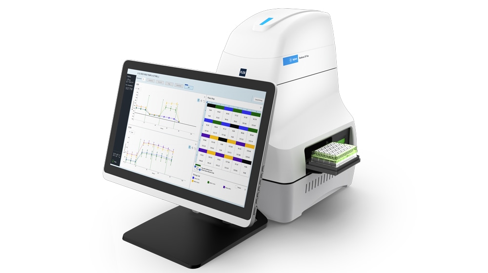
Seahorse XF Flex Analyzer
The Agilent Seahorse XF Flex analyzer measures and reports the oxygen consumption rate (OCR), proton efflux rate (PER) or extracellular acidification rate (ECAR), as well as ATP production rates in a 24-well format. It offers optimized workflows and dedicated consumables to enable real-time metabolic analysis with 3D study models, such as organoids and tissue materials. It also provides wide signal dynamic range through the use of dedicated consumables. Mitochondrial respiration and glycolytic activity are critical attributes of energy metabolism and vital indicators of mitochondrial health, toxicity, glycolysis, and overall cellular (dys) function. The ability to analyze these parameters in more complex three-dimensional structures will reveal deeper insights and enhance the translatability of your research.
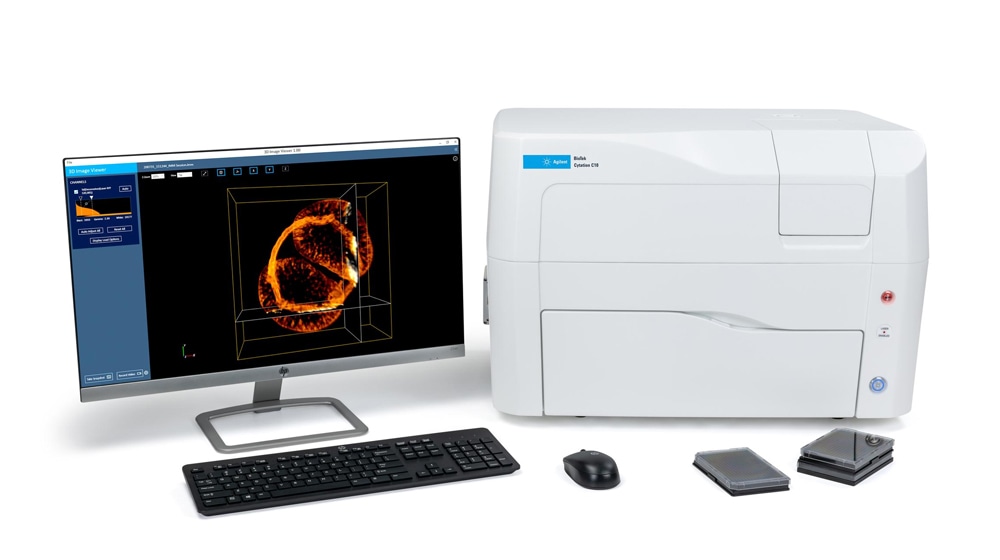
BioTek Cytation C10 Confocal Imaging Reader
The Agilent BioTek Cytation C10 unlocks detailed insights into complex biological systems. Featuring multiple, diverse imaging modes, it is uniquely designed for high-resolution tissue and whole-organism imaging. Its spinning disk confocal, paired with water immersion objectives, provides unparalleled clarity. Additionally, the Deep Sectioning Disk (DSD) allows for a clear view into thicker samples—a crucial capability for studying biomolecules and cellular structures within 3D cultures, tissues such as the mouse retina, and intact zebrafish embryos.
Webinar OnDemand

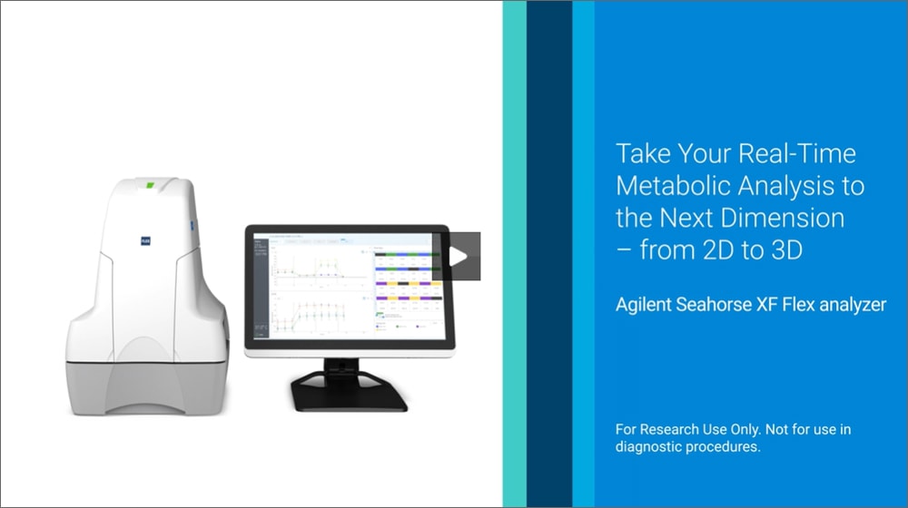
The New Agilent Seahorse XF Flex Analyzer Takes Real Time Metabolic Analysis to the Third Dimension
Three-dimensional (3D) models offer significant benefits for biomedical research by providing a more realistic understanding of biological processes and diseases than traditional 2D methods. The webinar will present Agilent's latest innovation, the Seahorse XF Flex analyzer. This platform offers flexibility and an optimized workflow for functional metabolic studies in 3D models, including ex vivo tissue samples and organoids, pushing the boundaries of metabolic research.
Learning objectives:
- An introduction to the enhanced metabolic assay platform developed for applications using 3D study models.
- Techniques for optimizing assay performance with different sample types and data normalization options
- Guidance on selecting the appropriate consumables to ensure successful results
- Examples of how improved sensitivity and precision will help you to achieve high-quality data
Featured Video
Seahorse XF Flex analyzer and 3D applications
The 24-well Agilent Seahorse XF Flex analyzer features improved sensitivity and precision, a broad detection range, and dedicated consumables designed for optimized workflows compatible with 3D study models, such as tissue, organoids and coculture materials.
Additional Resources
- 3D Metabolic Analysis: From 2D to Translatable Research
- Histology and Whole Organism Imaging
- Agilent Seahorse XF Applications in Cell Metabolism
- Agilent Seahorse XF 3D Mito Stress Test Tissue Workflow
- Histology and Slide Scanning - TekTalk Newsletter
- Zebrafish Applications - TekTalk Newsletter
For Research Use Only. Not for use in diagnostic procedures.
RA250701.422