BioTek Gen5 Software for Imaging & Microscopy
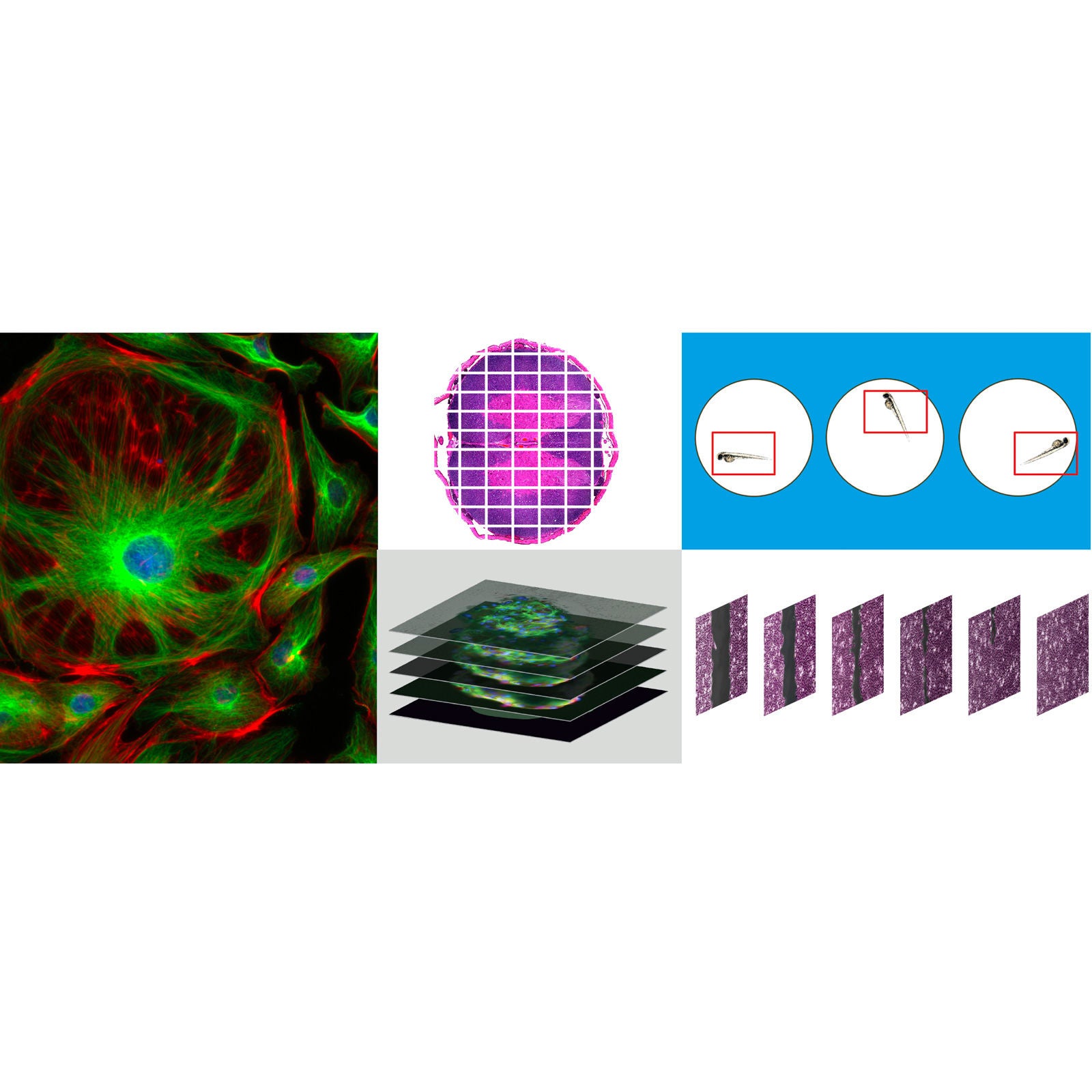
Transform images into biological insight.
- Agilent BioTek Gen5 software for imaging and microscopy combines instrument control, automated image acquisition, image processing, and cellular analysis in one intuitive interface. Define widefield and confocal workflows for 2D and 3D samples using tools such as montaging, Z-stacking, autofocus, Z-projection, deconvolution, and 3D rendering. Advanced analysis capabilities support multilevel masking, measurements across imaging channels, and detailed cell morphology assessments. Optional analysis modules further extend functionality for specialized workflows, including AI-based cell identification and segmentation to help reduce threshold setting and iterative parameter tuning for cell counting, confluence, and morphology analysis.
- Integrated workflow: Control instruments, acquire images, process data, and perform analysis in one interface.
- Flexible image acquisition: Automate montaging, Z-stacking, autofocus, and kinetic imaging.
- Advanced image processing: Apply background removal, deconvolution, stitching, Z-projection, and 3D rendering.
- Expanded cellular analysis: Use multilevel masking, multichannel measurements, and object morphology metrics.
- Optional AI segmentation: Reduce threshold setting and parameter tuning for cell identification and analysis.
Discover more
Overview
Future-ready features
Automated image acquisition
Automate single-image, montage, Z-stack, autofocus, and kinetic acquisition with compatible BioTek imaging systems.
AI cell identification
Add AI-based segmentation to reduce threshold setting and iterative tuning for cell counting and confluence analysis.
Multilevel masking
Create linked masks to measure cellular compartments and quantify spatially resolved signals in complex assays.
Multichannel measurements
Measure signals from all imaging channels to characterize objects and cellular regions within a unified analysis.
2D and 3D image processing
Use stitching, deconvolution, Z-projection, and 3D rendering to prepare complex image sets for analysis.
Object-level analysis
Quantify cell count, confluence, size, shape, intensity, morphology, and other object-level measurements.
Specialized analysis modules
Extend Gen5 with modules for neurite outgrowth, spot counting, object tracking, AutoROI, and scratch assays.
Data visualization and export
Create graphs, scatterplots, and histograms, then export data, images, and movies for downstream use.
How it works
From acquisition to analysis
Gen5 brings image acquisition, processing, analysis, and visualization together in one environment. Define protocols, capture images automatically, apply processing steps, quantify results, and prepare outputs without moving between multiple software packages.


Prepare images for analysis
Integrated processing tools prepare images for quantitative analysis. Background correction, deconvolution, stitching, Z-projection, 3D rendering, and kinetic image alignment support workflows from routine imaging to complex 3D samples.
Quantify complex image data
Object-based analysis supports confluence, cell counting, morphology, and cellular or subcellular measurements. Optional modules extend Gen5 for AI-based cell identification, object tracking, spot counting, neurite outgrowth, scratch assays, and AutoROI.

Applications
Advanced Gen5 analysis modularity
Available application-specific software modules for Gen5 enable advanced, automated analyses. Modules include neurite outgrowth, spot counting, automatic region of interest identification, single object tracking, and kinetic scratch wound healing assays.


Image and data publishing
Higher-level graphical representations of calculated results can also be created within Gen5 software, including IC50/EC50 graphs, linear and parallel line analysis, scatter plots, and histograms. Graphs, data, and high resolution-images and movie files can then be automatically exported from the software to be used for publishing or other downstream applications.
Technology
Object tracking
With the incorporation of the object tracking module, available within Agilent BioTek Gen5 software for imaging and microscopy, this can be easily accomplished in a fully automated fashion.
Automatic region of interest identification
This process enables walk-away slide scanning for four or more slides, if combined with Agilent automation offerings, and can be used with the Agilent BioTek Lionheart automated microscopes and Agilent BioTek Cytation imaging multimode readers.
Spot counting
The spot counting module available in Agilent BioTek Gen5 software enables the number of spots counted, and other information, to be tied to the original primary mask data, allowing these values to be known for each object initially identified in the image.
Neurite outgrowth
Neurite outgrowth is the process by which new projections form on neurons. Neuronal outgrowth is important in the study of normal development and regeneration, and the study of how inhibition or stimulation of neurite outgrowth occurs in disorders such as Parkinson’s disease and Alzheimer’s disease.
Tools

Customer reviews for BioTek Gen5 Software for Imaging & Microscopy
Explore customer reviews of this product or write your own review


Agilent Cell Analysis Webinars
Featured webinars help you stay informed about timely cell analysis research topics.

One system, any live-cell assay
The BioTek Cytation is ready for any assay and any scientific demand. Explore each pillar driving discovery.
Training & events
Cytation C10: An Affordable Confocal Imaging Reader ...
Cytation C10 Confocal Imaging Reader combines automated digital confocal and widefield microscopy with conventional multi-mode microplate reading.
Next Generation Cytation Technology: A ...
Our next generation Cytation optics facilitates a diverse array of workflows ranging from 3D cell culture assays through to whole organism imaging.
Multimodal zebrafish workflows on a single ...
Join us in this webinar to explore multimodal zebrafish applications that enable automated zebrafish workflows on a single instrument.
Related products
PRDT_1623188
PRDT_1623190
PRDT_1775872
PRDT_9809506
PRDT_1623200
PRDT_1623215
PRDT_1623188
PRDT_1623190
PRDT_1775872
PRDT_9809506
PRDT_1623200


