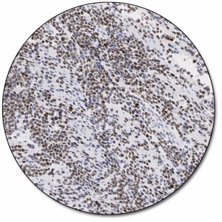
Ki-67 Antigen (Dako Omnis)

- Monoclonal Mouse Anti-Human

No data to display
This product is not available for purchase by the general public.
Product Details
Features
- Optimized staining performance of both high and low-expression structures
- Dako Omnis and the dynamic gap staining technology provides a consistent, uniform staining with excellent morphology
- Crisp and clear staining with no background
- Optimal laboratory efficiency with ready-to-use antibodies on Dako Omnis
- The staining performance of all FLEX RTU antibodies has been defined, tested and approved through collaboration with leading, international pathology experts.
Specifications
| Application |
|
| Clone |
|
| Code Number |
|
| Immunogen |
|
| Isotype |
|
| Reagent Provided |
|
| Solutions |
|
| Species |
|
| Specificity |
|
Literature
- Technical Overviews
-
Immunohistochemical Staining Methods - Education Guide, Sixth Edition
The focus of the sixth edition of the IHC Guidebook is to provide a comprehensive immunohistochemistry (IHC) resource for lab managers, lab technicians, learning pathologists, and students from around the world.
- Technical Overviews
- English
- 30 Nov 2021
- 27.05 MB
Support
- Instructions for Use
-
Featured References
- Product Description References
-
1. Gerdes J, Becker MH, Key G, Cattoretti G. Immunohistological detection of tumour growth fraction (Ki-67 antigen) in formalin-fixed and routinely processed tissues. J Pathol 1992,168:85-6.
2. Cattoretti G, Becker MH, Key G, Duchrow M, Schlüter C, Galle J, et al. Monoclonal antibodies against recombinant parts of the Ki-67 antigen (MIB 1 and MIB 3) detect proliferating cells in microwave-processed formalin-fixed paraffin sections. J Pathol 1992,168:357-63.
3. Scholzen T, Gerdes J. The Ki-67 protein: from the known and the unknown [review]. J Cell Physiol 2000,182:311-22.
Promotions