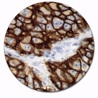
Epithelial antigen is a transmembrane glycoprotein functioning as a cellular adhesion molecule. This epithelium-specific antigen is broadly distributed in epithelial cells, and displays a highly conserved expression in carcinomas. The antibody is a useful aid for classification of adenocarcinoma. Anti-Epithelial Antigen may also aid in classification of micrometastases in lymph nodes of esophageal carcinoma as well as in classification of basal and squamous cell carcinomas of the skin.
This product is not available for purchase by the general public.
Product Details
Features
Optimized staining performance of both high and low-expression structures
Crisp and clear staining with no background
Optimal laboratory efficiency with ready-to-use antibodies on Autostainer Link 48
The staining performance of all antibodies has been defined, tested and approved through collaboration with leading international pathologists. Check the Atlas of Stains - a guide to diagnostic accuracy.
Specifications
Application
Formalin
HIER
Clone
Ber-EP4
Code Number
IR637
Immunogen
MCF-7 cells (human breast carcinoma cell line) (4). See package insert for reference(s).
Isotype
IgG1, kappa
Reagent Provided
Ready-to-use monoclonal mouse antibody provided in liquid form in a buffer containing stabilizing protein and 0.015 mol/L sodium azide.
Solutions
Autostainer Link 48
Species
Mouse Anti-Human
Specificity
Analysis of immunocomplexes between the antibody and lysate of 125I surface-labeled MCF-7 cells in SDS-PAGE under reducing conditions shows that the antibody labels two polypeptides of 34 kDa and 39 kDa, respectively, corresponding to epithelial antigen. Under non-reducing conditions, the polypeptides appear as 39 kDa and 41 kDa, while deglycosylation reduces the size to 31 kDa, and 36 kDa. In immunoprecipitation experiments, the Ber-EP4 antibody blocks the reaction of the HEA125 antibody with MCF-7 cell lysate and vice versa, showing that the two antibodies react with the same antigen. The two antibodies also produce identical staining results in cells and tissues (4). Of 37 cell lines tested, the antibody homogeneously labels all (10/10) carcinoma cell lines, whereas all non-epithelial cell lines (26/27) are not labeled except for the erythromyeloid cell line K562 (4). See package insert for reference(s).