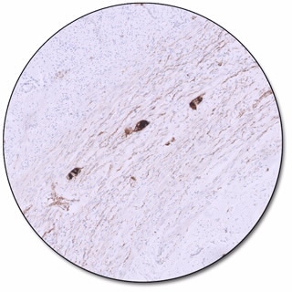
Normal appendix
Calretinin (Dako Omnis)
IVD

For In Vitro Diagnostic Use.
- Monoclonal Mouse Anti-Human

No data to display
This product is not available for purchase by the general public.
Product Details
Features
- Optimized staining performance of both high and low-expression structures
- Crisp and clear staining with no background
- Optimal laboratory efficiency with ready-to-use antibodies on Dako Omnis family of instruments
- Consistent, uniform staining with excellent morphology enabled by dynamic gap staining technology on the Dako Omnis family of instruments
- Defined, tested, and approved staining performance for all FLEX RTU antibodies through collaboration with leading international pathology experts
Specifications
| Application |
|
| Clone |
|
| Code Number |
|
| Form/Configuration |
|
| Immunogen |
|
| Isotype |
|
| Reagent Provided |
|
| Solutions |
|
| Specialty |
|
| Species |
|
| Specificity |
|
| Volume |
|
Literature
- Catalogs
-
Atlas of Controls
The Atlas of Controls illustrates the staining performance of each antibody in the FLEX Ready-to-Use system.
- Catalogs
- English
- 31 Jul 2026
- 37.96 MB
- Technical Overviews
Support
- Instructions for Use
-
Featured References
- Product Description References
-
1.Rogers, J. H. Calretinin: A gene for a novel calcium-binding protein expressed principally in neurons. J. Cell Biol. 1987, 105 (3), 1343–1353. DOI: 10.1083/jcb.105.3.1343.
2.Pochet, R.; Blachier, F.; Malaisse, W.; Parmentier, M.; Pasteels, B.; Pohl, V.; Résibois, A.; Rogers, J.; Roman, A. Calbindin-D28 in mammalian brain, retina, and endocrine pancreas: immunohistochemical comparison with calretinin. In Calcium Protein Signaling; Hidaka, H.; Carafoli, E.; Means, A. R.; Tanaka, T., Eds.; Adv. Exp. Med. Biol.; Springer: Boston, MA, 1989; Vol. 255, pp 435–443. DOI: 10.1007/978-1-4684-5679-0_46.
3.Bertschy, S.; Genton, C. Y.; Gotzos, V. Selective immunocytochemical localisation of calretinin in the human ovary. Histochem. Cell Biol. 1998, 109 (1), 59–66. DOI: 10.1007/s004180050202.
4.Lukas, W.; Jones, K. A. Cortical neurons containing calretinin are selectively resistant to calcium overload and excitotoxicity in vitro. Neuroscience 1994, 61 (2), 307–316. DOI: 10.1016/0306-4522(94)90233-X.
5.Oyama, Y.; Hamasaki, M.; Matsumoto, S.; Sato, A.; Tsujimura, T.; Nabeshima, K. Short 57 kb CDKN2A FISH probe effectively detects short homozygous deletion of the 9p21 locus in malignant pleural mesothelioma. Oncol. Lett. 2021, 22 (6), 813. DOI: 10.3892/ol.2021.13074.
6.Taheri, Z. M.; Mehrafza, M.; Mohammadi, F.; Khoddami, M.; Bahadori, M.; Masjedi, M. R. The diagnostic value of Ki-67 and repp86 in distinguishing between benign and malignant mesothelial proliferations. Arch. Pathol. Lab. Med. 2008, 132 (4), 694–697. DOI: 10.5858/2008-132-694-TDVOKA.
7.Kaur, G.; Nijhawan, R.; Gupta, N.; Singh, N.; Rajwanshi, A. Pleural fluid cytology samples in cases of suspected lung cancer: An experience from a tertiary care centre. Diagn. Cytopathol. 2017, 45 (3), 195–201. DOI: 10.1002/dc.23659.
8.Kim, J.-H.; Kim, G.-E.; Choi, Y. D.; Lee, J. S.; Lee, J. H.; Nam, J.-H.; Choi, C. Immunocytochemical panel for distinguishing between adenocarcinomas and reactive mesothelial cells in effusion cell blocks. Diagn. Cytopathol. 2009, 37 (4), 258–261. DOI: 10.1002/dc.20986.
9.Naka, T.; Hatanaka, Y.; Tabata, Y.; Takasawa, A.; Akiyama, H.; Hida, Y.; Okada, H.; Hatanaka, K. C.; Mitsuhashi, T.; Kushitani, K.; et al. Identification of novel diagnostic markers for malignant pleural mesothelioma using a reverse translational approach based on a rare synchronous tumor. Diagnostics 2022, 12 (2), 316. DOI: 10.3390/diagnostics12020316.
Related Products
Promotions