Detailed Characterization of Cell Health and Morphology
May 2026
A comprehensive understanding of the physiological state or health of your cells is vitally important for achieving reliable experimental outcomes, as results are highly dependent on the initial condition of the cells. Commonly investigated aspects of cell health include cell stress, apoptosis, necrosis, autophagy, metabolic phenotype, and morphological alterations.
Traditional biochemical and imaging assays typically provide only endpoint measurements, lacking the biological and temporal context needed to fully understand dynamic cellular processes. To address these limitations, Agilent offers a broad portfolio of live cell–based solutions designed to capture cell morphology, function, and behavior over time.
By combining complementary technologies such as high content live cell imaging with the Agilent BioTek Cytation 9 cell imaging multimode reader, real time metabolic analysis with Agilent Seahorse XF technology, and label free kinetic monitoring with Agilent xCELLigence RTCA, researchers can assess cell health from multiple perspectives. This integrated approach enables scientists to determine not just whether cells are viable, but how they function over time.
Together, these platforms unify imaging, functional measurements, and real time kinetic analysis to deliver both rich phenotypic insight and quantitative robustness. Whether characterizing the metabolic signatures of immune cell activation or monitoring the long-term structural integrity of complex 3D organoids, this flexible ecosystem empowers laboratories to move beyond static endpoint snapshots and capture the full kinetic narrative of cellular health and disease.
Featured Application Notes
Automated Kinetic Imaging Assay of Cell Proliferation in 384-Well Format
Kinetic tracking of live cells is vital for accurate cell death quantification. This 72-hour study reveals that using absolute fluorescent apoptosis counts skews EC50 values due to ongoing cell proliferation. Utilizing the Cytation to capture whole wells allows researchers to normalize fluorescent signals against label-free total cell counts. This critical morphological normalization eliminates bias, revealing true dose-response metrics and precise onset times for varying cell death mechanisms.

High-Throughput Platform for Fluorescence Image-Based Neurite Outgrowth Analysis
Quantifying neuronal morphology is crucial for neurobiology research. This study evaluates neurite outgrowth in human iPSC-derived and primary mouse neurons exposed to known modulators. By tracking structural metrics like total neurite length, branching, and soma counts, distinct dose-dependent responses were uncovered. For instance, triptolide significantly inhibited neurite complexity in human iPSCs. The Cytation combined with Gen5 software seamlessly automated this complex multidimensional analysis.
Tek Tips
Use Kinetic Imaging to Resolve Reversible Stress from Irreversible Cell Death
Why it matters
Endpoint viability assays often confound growth arrest, adaptive stress, and committed cell death—an issue amplified in 3D cultures and organoids due to spatial heterogeneity and asynchronous responses (1). These limitations are well recognized in NAM frameworks, where mechanistic confidence and reproducibility are emphasized over single endpoint measurements (3).
Best practice guidance
- Prioritize kinetic, image based measurements that track proliferation alongside early apoptosis and loss of membrane integrity, rather than single time point readouts (2).
- Separate early and late death events to distinguish reversible stress from irreversible loss of viability, particularly for delayed or gradual responses.
- Normalize functional signals to total cell or structure counts to control for condition dependent growth differences—an approach aligned with NAM recommendations for objective assay acceptance criteria (1,2).
- In 2D adherent cultures, impedance can provide a complementary, label free functional readout of attachment and barrier integrity, while imaging supplies the morphological and biological context needed for interpretation.
QC relevance
- Distinct kinetic patterns (cytostasis, delayed apoptosis, rapid lysis) support mechanism aware QC checks increasingly expected in NAM compatible workflows (2).
- In joint imaging–impedance workflows, impedance may flag early functional change in 2D, while imaging confirms biological relevance; for 3D systems, imaging remains essential (1).
Define QC Metrics as Release Criteria for 3D Viability Assays
Why it matters
Organoid assays show intrinsic variability in size, morphology, and growth. Without explicit QC gates, this variability can mask treatment effects and undermine reproducibility—key concerns driving the adoption of NAM and organoid standardization efforts (1,3).
Best practice guidance
- Apply non invasive imaging QC early to verify consistent structure formation and growth before treatment, consistent with hierarchical QC strategies proposed for NAM relevant 3D systems (1,2,3).
- Track object level metrics (count, size, morphology) alongside viability signals to contextualize responses and reduce false calls (3).
- Use longitudinal measurements to ensure changes reflect true biological effects, not delayed signal development.
- Where parallel 2D controls are included, impedance may provide supportive functional context; however, imaging derived metrics should define acceptance and release criteria for 3D assays.
QC relevance
- Imaging derived metrics can function as pre analysis release criteria, a practice explicitly aligned with NAM guidance calling for predefined, objective assay acceptance rules (1,2).
- A hierarchical QC strategy—imaging as the primary gate, with orthogonal 2D functional checks where appropriate—supports scalable, regulatorily relevant workflows (1,2).
References
- H. Castiglione, L. Madrange, C. Baquerre, B. G. C. Maisonneuve, T. Lemonnier, J. P. Deslys, et al. Towards a quality control framework for cerebral cortical organoids. Sci Rep 2025,15, 1, 29431.
- OECD, Guidance on Good In Vitro Method Practices (GIVIMP), 2018 – NAM aligned requirements for predefined acceptance/rejection criteria
- H. Lee, S. Yang, K. J. Lee, S. N. Kim, J. S. Jeong, K. Y. Kim, et al. Standardization and quality assessment for human intestinal organoids. Front Cell Dev Biol 2024, 12, 1383893.
Product Spotlights
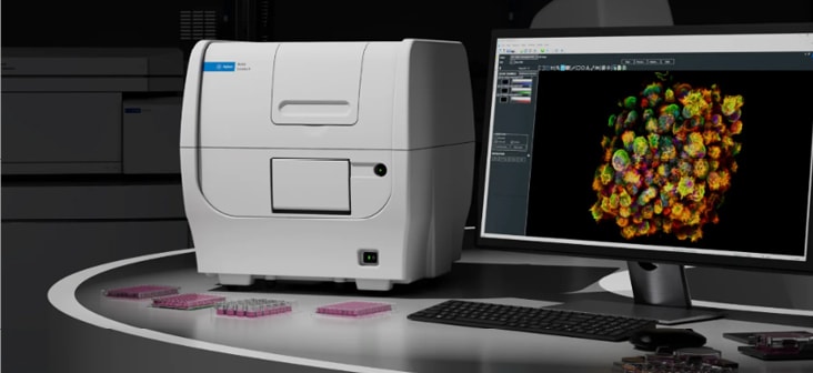
BioTek Cytation 9 Cell Imaging Multimode Reader
The BioTek Cytation 9 offers flexible, high-resolution analysis of cell health and morphology. Whether analyzing the intricate dendritic branching of neurons to the dense 3D architecture of organoids, the platform delivers the phenotypic clarity needed to track cellular fitness over time. Automated Z-stacking transforms thick biological samples into fully focused insights, while precise environmental controls maintain the stability required to capture the kinetic narrative of sensitive live-cell models. By bridging the gap between structural morphology and functional readouts, the Cytation 9 provides a comprehensive view of cellular fitness.
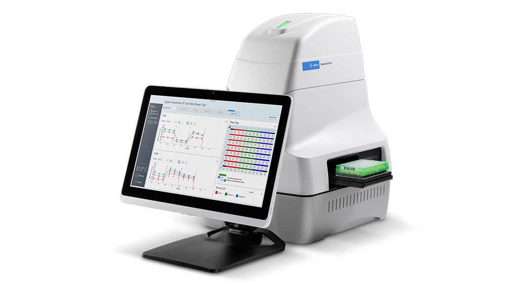
Seahorse XF Analyzers
Seahorse XF technology enables the real-time measurement of cellular bioenergetics by simultaneously quantifying oxygen consumption rate (OCR) and extracellular acidification rate (ECAR). This provides direct insight into mitochondrial respiration and glycolytic activity, revealing cellular stress, activation, toxicity, or dysfunction, often before detectable morphological alterations enabling early functional characterization of cell health. Seahorse XF is widely used to assess mitochondrial health and metabolic flexibility across 2D and 3D models, supporting applications in disease modeling, drug screening, and cell quality assessment.
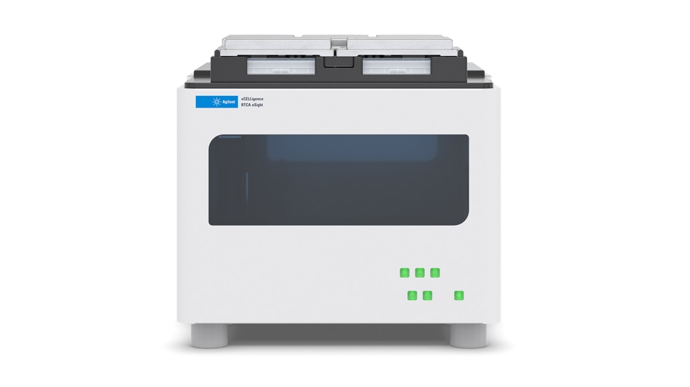
xCELLigence RTCA Technology
xCELLigence Real‑Time Cell Analysis (RTCA) systems provide continuous, label‑free, noninvasive monitoring of cell health using impedance‑based Cell Index (CI) measurements. Signals are captured via gold microelectrodes embedded in E‑Plate microtiter plates. As cells attach, spread, proliferate, and undergo morphological changes, dynamic changes in impedance enable uninterrupted monitoring. Each cell line exhibits a distinctive CI profile, deviations from which provide immediate insights into changes in cell health, enabling early detection of cytotoxicity, stress, or functional alterations.
Webinars On Demand
Agilent BioTek Cytation 9 Technology: Orchestrating Orthogonal Cell Analysis at Scale
In modern research, understanding biology demands that we connect diverse data types: kinetic, spatial, and functional. Agilent BioTek Cytation 9 technology empowers you to bridge these dimensions across your research program. By combining advanced quantitative imaging with multimode detection, it enables you to investigate cellular and biomolecular processes from multiple angles, when and where it matters most.
This webinar will showcase how you can expand your imaging capabilities across additional channels while seamlessly integrating rapid, multimode detection. With refined environmental control and faster acquisition speeds, Cytation 9 supports studies ranging from live-cell dynamics to mechanistic investigation, allowing you to align pathway-level signals with cell-state transitions over time.
We’ll present case studies spanning a range of applications, from rapid plate-based kinetic measurements to high-resolution imaging and protein-protein interaction assays. Whether you’re analyzing proliferation, migration, complex 3D models, or studying signaling pathways, you’ll see how orthogonal approaches strengthen your conclusions.
Additional Resources
Application & Industries
Application Notes
- Real-Time Quality Control and Functional Assessment of Mesenchymal Stem Cells for Cellular Therapies
- Stimulation of Human Peripheral Blood Mononuclear Cells
- Automated, Label-Free, Kinetic Neurite Outgrowth Analysis
- Automated Cell Viability Assay
- Use of Phase Contrast Imaging to Track Morphological Cellular Changes due to Apoptotic Activity