Vitality hrGFP Mammalian Expression Vectors - Details & Specifications
The green fluorescent protein (GFP) has become an extremely versatile tool for tracking and quantifying biological entities in the fields of biochemistry, molecular and cell biology, as well as high throughput screening and gene discovery. GFPs have been identified in a wide range of coelenterates, and while recently the number of cloned GFPs has expanded, to date the best characterized proteins are those from the jellyfish Aequorea victoria and the anthazoan Renilla reniformis.
We have isolated a cDNA clone for the R. reniformis GFP, and have fully humanized the gene using codons preferred in highly expressed human genes. The humanized recombinant GFP (hrGFP) cDNA was then subjected to random mutagenesis to generate the brighter variant, hrGFP II. The fluorescence spectra for hrGFP and hrGFP II are essentially identical to the published spectrum for the purified native protein, with the major excitation peak at 500 nm and the emission peak at 506 nm.
We have expressed the hrGFP II in a wide range of human, rodent, and simian cell lines (see Figures 1 and 2), and observed levels of fluorescence higher than that for the red-shifted, humanized variant of Aequorea GFP (EGFP) in all cell-types tested (see Figure 3). In addition, stable GFP-expressing cell lines are produced much more efficiently using the Agilent hrGFP compared with EGFP, since Aequorea GFP is often cytotoxic when expressed at low levels.
Agilent offers four Vitality hrGFP II vectors for mammalian expression. These vectors support a variety of expression configurations, thus providing ideal expression options for each specific application. The hrGFP II allows expressed genes to be easily visualized using fluorescence microscopy or fluorescence-activated cell sorting (FACS). We also offer a Vitality full-length hrGFP polyclonal antibody (Catalog #240141 and #240142) which recognizes the wild type green fluorescent protein, hrGFP, and the hrGFP II mutant as well as N- and C- terminal fusions to these proteins. Applications include Western blotting, immunoprecipitation, and flow cytometry. Because hrGFP is derived from a different organism, the hrGFP antibody does not cross react with the Aequorea victoria GFP variant, EGFP.
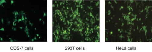
Figure 1: The Renilla reniformis humanized recombinant green fluorescent protein II (hrGFP II) was expressed in a range of mammalian cell types by transfection with the phrGFP II-C vector. Cells were plated at 2.4–6 × 10⁵ cells per well in 6-well plates the day before transfection. Transfection was performed using a cationic lipid reagent at DNA:reagent (w/v) ratios of 1:1.25 (HeLa) and 1:2 (COS-7 and 293T) and incubated for 5 hours. All images were taken with a 10-second exposure time.
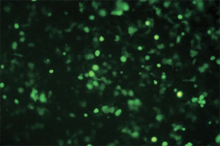
Figure 2: The Renilla reniformis humanized recombinant green fluorescent protein II (hrGFP II) was expressed in 293T cells by transfection with the pIRES-hrGFP II vector. Cells were plated at 6 × 10⁵ cells per well in a 6-well plate the day before transfection. Transfection was performed using a cationic lipid reagent at DNA:reagent (w/v) ratio of 1:2 and incubated for 5 hours. The image was taken 48 hours post transfection with a 10-second exposure time.
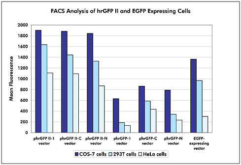
Figure 3: FACS analysis of reporter brightness. Transient transfections of COS-7, 293T, and HeLa cells with various GFP-expressing vectors were performed using a cationic lipid reagent at DNA:reagent (w/v) ratios of 1:1.25 (HeLa) and 1:2.5 (COS-7 and 293T). The cells were collected at 24 hours post-transfection for FACS analysis. Mean fluorescence is defined by the total fluorescence in the marker region (M1) divided by the total cells collected in the marker region (M1).
For Research Use Only. Not for use in diagnostic procedures