StrataClone Mammalian Expression Vector Systems - Details & Specifications
The StrataClone Mammalian Expression Vectors are designed for high-level expression of epitope-tagged proteins in mammalian cells. Use of StrataClone technology for the cloning steps allows rapid 5-minute cloning of blunt-ended PCR products, produced using high-fidelity PCR enzymes, into the epitope tagging/expression vectors. The StrataClone cloning method is summarized in Figure 1 and discussed in greater detail below.
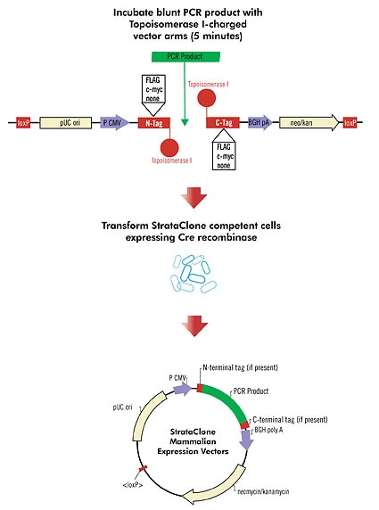
Figure 1: Overview of the StrataClone mammalian expression cloning method. Five varieties of vector arms are available, with either an N-terminal tag (FLAG® or c-myc), a C-terminal tag (FLAG or c-myc), or no tag.
The epitope tagging technique involves fusion of a protein of interest to a peptide epitope that is recognized by a readily available antibody. With this technique, expression of the fusion protein is monitored using a tag-specific antibody, allowing a new protein to be studied without generating a new antibody specific to the protein of interest. Epitope tagging can be used to localize gene products in living cells, identify associated proteins, track the movement of fusion proteins within the cell, or characterize new proteins by immunoprecipitation.
The StrataClone Mammalian Expression Vectors contain sequences encoding three copies of either the FLAG® or the c-myc epitope at either the N or C terminus. These specific epitope tags are small, highly immunoreactive, and are not likely to interfere with the function of the target protein. The synthetic FLAG epitope is composed of eight amino acid residues (DYKDDDDK). The c-myc epitope is derived from the human c-myc gene and contains ten amino acid residues (EQKLISEEDL). Tagged constructs generated in the StrataClone Mammalian Expression Vectors can be transfected into mammalian cells and the fusion protein can be easily characterized using commercially available antibodies. The presence of three copies of the epitope in each vector enhances detection of the fusion protein in downstream applications. An expression vector that lacks epitope tagging sequences is also available to allow expression of untagged protein or fusion proteins with a custom tag encoded by the PCR insert. In addition to the epitope tag sequences, the StrataClone Mammalian Expression Vectors contain features for expression of fusion proteins in eukaryotic cells. The cytomegalovirus (CMV) promoter allows constitutive expression of the cloned DNA in a wide variety of mammalian cell lines. The vectors contain a neomycin-resistance gene under control of a mammalian promoter for selection in mammalian cells.
Using the method summarized in Figure 1, StrataClone blunt PCR cloning technology exploits the combined activities of topoisomerase I from Vaccinia virus and Cre recombinase from bacteriophage P1. In vivo, DNA topoisomerase I assists in DNA replication by relaxing and rejoining DNA strands. Topoisomerase I cleaves the phosphodiester backbone of a DNA strand after the sequence 5´-CCCTT, forming a covalent DNA–enzyme intermediate which conserves bond energy to be used for religating the cleaved DNA back to the original strand. Once the covalent DNA–enzyme intermediate is formed, the religation reaction can also occur with a heterologous DNA acceptor. The Cre recombinase enzyme catalyzes recombination between two loxP recognition sequences.
Each StrataClone PCR cloning vector mix contains two blunt-ended DNA arms. Both arms are charged with topoisomerase I on one end and contain a loxP recognition sequence on the other end. Blunt-ended PCR products, produced by proofreading PCR enzymes, are efficiently ligated to these vector arms in a 5-minute ligation reaction by topoisomerase I-mediated strand ligation.The resulting linear molecule (vector armori–PCR product–vector armneo/kan) is then transformed, with no clean-up steps required, into a competent cell line engineered to transiently express Cre recombinase. Cre-mediated recombination between the vector loxP sites creates a circular DNA molecule that is proficient for replication in E. coli cells growing on media containing kanamycin. Maps for the circular plasmid products of recombination are shown in Figures 2–6. The circular maps provided are for reference only; actual circular DNA plasmids will contain user-specific PCR product inserts.
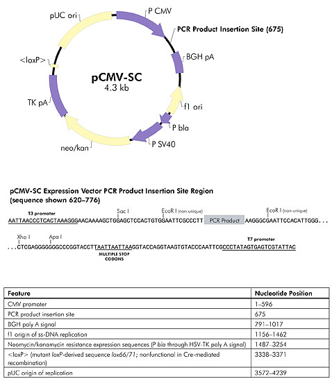
Figure 2: StrataClone blunt PCR cloning vector pCMV-SC. The circular map shown represents the product of topoisomerase I-mediated ligation of the supplied vector arms to a PCR product of interest followed by Cre-mediated recombination.
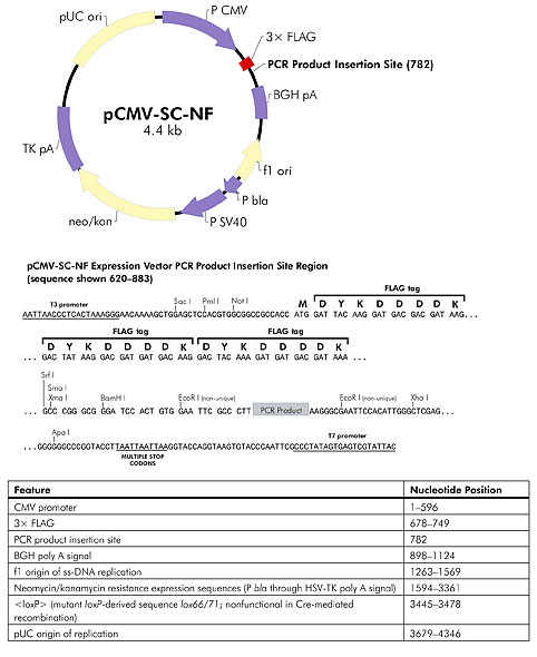
Figure 3: StrataClone blunt PCR cloning vector pCMV-SC-NF. The circular map shown represents the product of topoisomerase I-mediated ligation of the supplied vector arms to a PCR product of interest followed by Cre-mediated recombination.
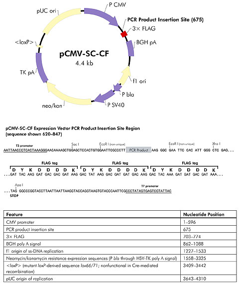
Figure 4: StrataClone blunt PCR cloning vector pCMV-SC-CF. The circular map shown represents the product of topoisomerase I-mediated ligation of the supplied vector arms to a PCR product of interest followed by Cre-mediated recombination.
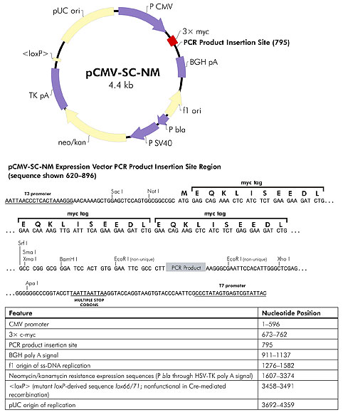
Figure 5: StrataClone blunt PCR cloning vector pCMV-SC-NM. The circular map shown represents the product of topoisomerase I-mediated ligation of the supplied vector arms to a PCR product of interest followed by Cre-mediated recombination.
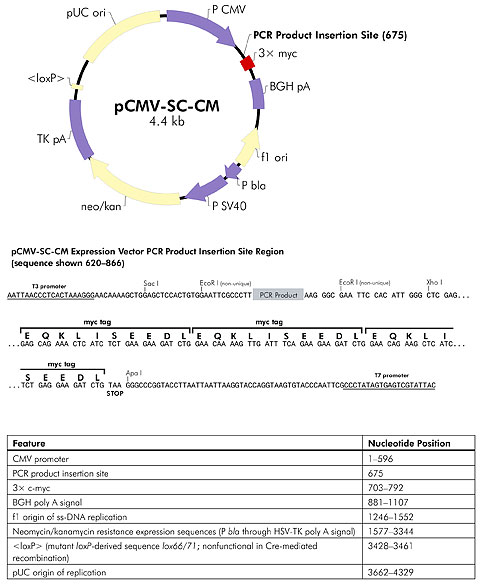
Figure 6: StrataClone blunt PCR cloning vector pCMV-SC-CM. The circular map shown represents the product of topoisomerase I-mediated ligation of the supplied vector arms to a PCR product of interest followed by Cre-mediated recombination.
For Research Use Only. Not for use in diagnostic procedures