Access Agilent eNewsletter, January 2014
>> Update My Profile | Subscribe to Access Agilent | Article Directory

Size Exclusion Chromatography: Excerpt from our newest “how to” guide
By Andrew Coffey
Agilent LC Applications Specialist
 Enlarge
Figure 1. Bio SEC mechanism.
 Enlarge
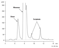
Figure 2. Intact MAb monomer and dimer separation using an Agilent Bio SEC-3 column.
Size exclusion chromatography (SEC) is frequently employed to separate and quantify biomolecules of different sizes. The mechanism of SEC is one of relatively slow diffusion into and out of the stagnant pool of mobile phase captured in the pores. Molecules that are too large to permeate into the pore structure will be excluded from the pores and quickly pass through the column. Smaller molecules will be able to penetrate the pores to various degrees depending on their size; with the smallest molecules diffusing furthest in the pore structure and eluting last (shown in Figure 1). SEC is also used to analyze the molecular weight distribution of polydisperse molecules, such as starches and other polysaccharides. SEC is particularly valuable in determining the degree of aggregation of therapeutic proteins where immunogenic responses can result and is often used during development and quality control, as shown in Figure 2.
For successful size exclusion chromatography, the sample should not interact with the sorbent contained within the column. Instead, the molecules should simply diffuse in and out of the stagnant pools of eluent trapped in the pores of the sorbent. Larger molecules cannot penetrate the pore structure easily, so they pass through the column more quickly than smaller molecules. This diffusion process means size exclusion chromatography is carried out using large column dimensions and much lower flow rates (linear velocities) than more common HPLC techniques.
There are a number of important factors to consider before choosing SEC. That’s why Agilent has introduced a new “How To” Guide to Size Exclusion Chromatography for Biomolecule Analysis. This handbook guides you through choosing the correct column, mobile phase, sample preparation and other key factors that can help you get the most from employing the SEC technique – a very useful reference for method development.
The right sample preparation techniques ensure the right results
It’s fundamental. Sample preparation techniques for SEC applications require that the properties of the sample being measured are never affected. As with all chromatographic techniques, it is essential to ensure that the sample does not contain particulate matter. This can be easily resolved by using a simple syringe filter, such as an Agilent Captiva PES Filter with 0.2- or 0.45-µm pore size. However, it is important to understand the nature of the particulate matter – filtering out the aggregates you are trying to measure is simply not going to work.
With size exclusion chromatography it is often necessary to inject larger volumes of more concentrated solutions in order to get sufficient detector response. This also can lead to changes in the composition of the sample, so care must be taken in your choice of sample preparation. For example, using sonication or elevated temperatures to dissolve proteins may actually induce aggregation through stress.
 Enlarge
Figure 3. SEC Method Development Guide.
Choose a column that includes all analytes of interest
Choosing the correct pore size is perhaps the most important factor for the size exclusion of biomolecules. It is important to ensure that all of the analytes of interest are able to penetrate the pore structure, at least to some extent. Choosing a column that excludes one or more components from the pore structure is simply not conducive to good chromatography. For most globular proteins, 300Å is a good choice and will cover many eventualities. It’s also good practice to employ a regular calibration with well-characterized standards (Figure 3). This will highlight any potential problems with your system or method.
Don’t let the mobile phase change the composition of your sample
For many biomolecules, an aqueous buffer containing phosphate and chloride – for example, a phosphate buffered saline (PBS) – will give the necessary balance between ensuring sample solubility and also ensuring a minimum of non-specific interactions between the analytes and the packing material. Since chloride ions are rather corrosive even to the stainless steel components of column hardware and instrument parts, a moderate strength phosphate buffer may be a better choice. Agilent recommends using 150 mM phosphate as eluent for many size exclusion applications, although most other salt additives are also suitable. As mentioned with sample preparation, it is important to ensure that the mobile phase does not change the composition of the sample under analysis.
Get the right biosystems for your bioapplications
There are, of course, many other factors that could affect your choice of column and your chosen analysis conditions. That’s why you’ll find all the detailed information you need to choose the right columns, systems, and supplies for conducting reliable and robust size exclusion chromatography in Agilent’s “How To” Guide to Size Exclusion Chromatography for Biomolecule Analysis.
From sample purification to analysis, Agilent biomolecule columns are easy to integrate into your workflow for a complete, reproducible, and high-quality solution. Biomolecules may be complex in structure, but their analysis is simplified by using Agilent biocolumns, systems, and supplies, including the Agilent 1260 Infinity Bio-inert LC system with a metal-free sample path and the Agilent 1290 Infinity LC. Each are designed to provide the highest speed, resolution, and ultra-sensitivity for UHPLC applications, including those using Agilent wide-pore 300Å ZORBAX 300StableBond columns with ultra-sensitivity for UHPLC analysis.
No matter your specific bioapplication, Agilent offers the tools and resources to help you conduct reliable, reproducible SEC analyses. So take a moment now and download our “How To” Guide to Size Exclusion Chromatography for Biomolecule Analysis to learn more.
>> Update My Profile | Subscribe to Access Agilent | Article Directory
Figure 2.

Conditions |
|
|---|---|
Column |
Agilent BioSEC-3, 7.8 x 300 mm, 3 µm |
Sample |
CHO-humanized MAb, 5 mg/mL – intact |
Eluent |
Sodium phosphate buffer, pH 7.0, 150 mM, 0 to 100% from 0 to 30 min |
Injection |
5 µL |
Temperature |
Ambient |
Flow rate |
1.0 mL/min |
Detector |
UV, 220 nm |
Intact MAb monomer and dimer separation using an Agilent Bio SEC-3 column.