Professor Philip Doble
Picture thisAgilent helps research director push boundaries of bioimagingPhilip Doble wants everyone to see how diseases progress and how they might be treated so he is developing new ways to create detailed 3-D images of affected tissues. Call it next-generation imaging. Doble directs the Elemental Bioimaging Facility at the University of Technology in Sydney, Australia, where he and his team are using the same equipment geologists use to look for metals within rocks, plus state-of-the-art ICP-MS instruments from Agilent.
A chemist by training, Doble knows that many proteins need metals to function properly. Metals like copper, zinc, and manganese. So he maps them along with all sorts of biological molecules thought to be important in disease. A key component of his work is constructing 3-D images from the brains of mice affected by various disorders. "That allows you to look at various neuroanatomical regions and how they relate to each other," Doble says. "You can look for what genes are expressed in various regions of the brain and try to determine what's going on from a mechanistic point of view. We try to do that with essential elements. Mapping them in three dimensions allows you to compare them against the neuroanatomical regions in the Allen Brain Atlas." The new technique begins with technology from the geology industry. "Some of our instruments were first developed to look at rocks for minerals and things. Essentially what we have is a laser that fires radiation at 193 or 213 nanometers at a tissue section that is 10 to 15 microns thick and it obliterates the tissue, turning it into little particles. These little particles are then swept from the cells into the Agilent ICP-MS—an absolutely fantastic instrument—where they are reduced to their constituent elements," Doble explains. "As you raster the laser across the tissue section, you get information about the spatial concentrations of elements at the point of laser sampling. So it's a timed result analysis. Once that's done, we take all that data and stick it back together much like the old dot-matrix printers assembled dots into letters." Doble believes he and his team are at the beginning of what will prove to be a powerful new technique in the field of metalomics and beyond. "The measurement of metals is an indirect measurement of metabolism because they are either concentrated or depleted depending on what's happening in the environment of the cells," Doble says. "For example, the transport proteins can tell you something about the progression of a disease and the effects of therapeutic intervention."
Doble, who has been collaborating with Agilent on various projects over the past 15 years, speaks highly of the company's instruments and support services ("I have nothing but good things to say"), and he credits Agilent with facilitating collaborations with other scientists. "We're imaging muscular dystrophy using this technology, in collaboration with researchers in the United States, and we've had some nice images from that of multiplexed proteins," he says. "By tagging various antibodies and multiplexing the tagging protocols against the various proteins in muscle sections, we could pick out which proteins were important using this technology." Some might say, "Why would you want to do that? We have other techniques like fluorescence." But Doble's next-generation imaging can augment that. "We're talking about micrometer resolution here, which allows us to look at a tissue section from a helicopter perspective as opposed to fluorescence, which looks at subcellular arrangements. Instead of being a replacement, our technique just gives you a whole lot more information," he says. "What we're moving toward is using virtual reality and gaming engines to try to put our data into an interactive environment so we can basically sit in a rocket ship and fly around these reconstructions. It's early days yet, but it may even be possible to link this to other multimodal images such as infrared spectroscopy or MRI as well."
At this point, Doble says, next-generation imaging is very much a research tool to work out the interplay between various transporter proteins, the environment, cofactors, and so on. "As we learn more about the way metabolism occurs, particularly in regard to metals, then this could potentially be used as a diagnostic. Once we've got the tagging protocols completely worked out, we should be able to do quantifiable biomarker imaging. Our hope is that this could be used to look at particular proteins on the surface of cells, so that you might then be able to say, 'Well, this particular tumor is susceptible to this particular drug. In terms of personalized medicine, it has a lot of potential applications."
|
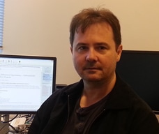
Philip Doble
Director of the Elemental Bioimaging Facility |
Selected publications
|
A guide to integrating immunohistochemistry and chemical imaging. Distributions of manganese in diverse human cancers provide insights into tumour radioresistance. Trehalose elevates brain zinc levels following controlled cortical impact in a mouse model of traumatic brain injury. Microfluidic high performance liquid chromatography-chip hyphenation to inductively coupled plasma-mass spectrometry. Imaging Metals in Brain Tissue by Laser Ablation - Inductively Coupled Plasma - Mass Spectrometry (LA-ICP-MS). |