Confocal Fluorescence Imaging
March 2021
Developed almost 70 years ago, confocal microscopy ushered in a major advancement to the field of fluorescence microscopy and has become a mainstay for imaging fluorescently labeled biological samples. Conventional widefield microscopy illuminates biological samples with a diffuse field of light that excites fluorophores above and below the focal plane of interest, resulting in a large amount of out-of-focus emitted light being captured by the system. By placing a pinhole in a conjugate focal plane between the sample and the light detector (camera or PMT), confocal microscopy blocks contaminating out-of-focus light, thus improving axial (z) resolution. This improved resolution means that clearer z-sections can be taken deeper within thick samples, such as spheroids and resected tissue, and three-dimensional biological relationships can be better elucidated.
There are two main designs of confocal microscopes with distinct features. In the first design, called Point Scanning (or Laser Scanning) Confocal, a single pinhole, a photomultiplier tube (PMT) detector, and a motorized mirror are employed. In this design, the motorized mirror directs excitation light emanating from a laser light source onto the specimen. The precision of the motorized mirror allows points across the sample to be sequentially illuminated, most commonly in a serpentine pattern. The emission light coming from each point as it is illuminated passes through the lone pinhole where out-of-focus light is blocked, and the remaining light is detected by the PMT. Once the scan is complete, the coordinates of each point of the scan area are mapped as individual pixels and used to reconstruct the final image. In contrast to point scanning (or laser scanning) confocal, in which a single pinhole, a photomultiplier tube (PMT) detector, and a motorized mirror are employed, spinning disk confocal employs a nipkow disk containing hundreds of pinholes organized in a spiralized array, which is placed in the conjugate focal plane. The disk is coupled to a motor which spins the disk at a set speed while excitation light is passed through the disk to illuminate the sample. Emission light from entire sample is then passed back through the pinholes of the spinning disk, where it is routed by a dichroic mirror and projected onto a camera chip (instead of a PMT). With the multiple pinhole design, spinning disk confocal can scan and illuminate the entire image area simultaneously, greatly reducing acquisition time and providing a gentler illumination method for labile fluorophores and live samples compared to laser scanning systems.
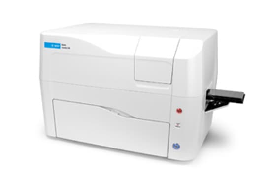
The Cytation C10 Confocal Imaging Reader uses the spinning disk design to combine automated confocal and widefield microscopy (including brightfield and phase contrast) with conventional multi-mode microplate reading in a unique, patented design. The Cytation C10 uses a Hamamatsu scientific CMOS (sCMOS) camera, Olympus objectives and laser-based illumination which enables excellent image quality. The new 3D viewer provides users with 3D rendering capabilities for optically sectioned samples captured with the confocal microscope. Its variable bandwidth monochromator-based multi-mode plate reading is based on the proven design of the market-leading BioTek Synergy products. Environmental controls enable live cell assays, and Gen5 software is specifically designed to make sample detection and image capture quick and effortless. The unique combination of features, supported applications, and affordable price point makes Cytation C10 a powerful benchtop workhorse for every laboratory.
Featured Applications
Optical Clearing for Improved Confocal Imaging of Thick Specimens
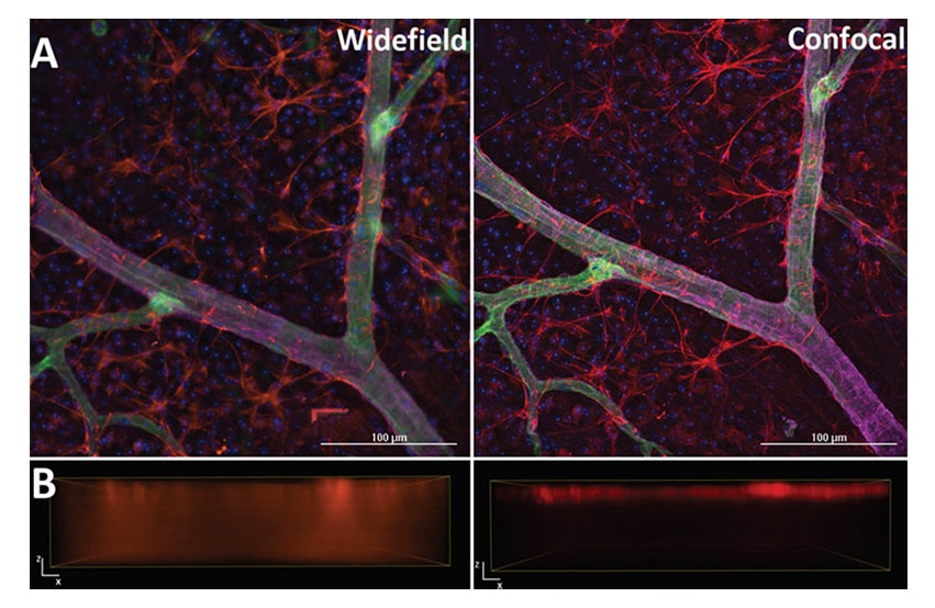
Developed almost 70 years ago, confocal microscopy has become a mainstay for imaging fluorescently labeled biological samples. Widefield microscopy illuminates biological samples with a diffuse field of light that excites fluorophores above and below the focal plane of interest, resulting in a blurry, less resolved image. By placing a pinhole in a conjugate focal plane between the sample and the light detector (camera or PMT), confocal microscopy blocks contaminating out-of-focus light, thus improving axial (z) resolution. This improved resolution means that finer z-sections can be taken deeper within thick samples, such as spheroids and resected tissue, and three dimensional biological relationships can be better elucidated.
Visualizing the Mouse Retinal Vasculature using the Cytation C10 Confocal Imaging Reader
The mouse retina is one of the most widely used in vivo systems to study angiogenesis and its associated pathologies. Mice are actually born without a retinal vasculature, however over the course of the first week of life, this vasculature expands radially from the optic opening to the periphery on the surface of the retina. At this expansion front are specialized tip endothelial cells that use actin-based cellular extensions called filopodia to sense growth factor gradients. These filopodia can vary in width by a few hundred nanometers. Once the initial vasculature reaches the periphery of the retina, nascent blood vessels start to branch into the retina, ultimately maturing into a tri-layer plexus.
Product Spotlight
Cytation C10 Confocal Imaging Reader
Cytation C10 Confocal Imaging Reader combines automated confocal and widefield microscopy with conventional multi-mode microplate reading. The spinning disk confocal module adds increased resolution and optical sectioning capabilities to the Cytation range. Cytation C10 also includes widefield fluorescence, brightfield and phase contrast optics. The multi-mode module has variable bandwidth monochromator-based optics for specificity and sensitivity. System control, image and data analysis provided by BioTek's Gen5 software.
Cytation C10 Confocal Imaging Reader
Cytation Cell Imaging Multi-Mode Plate Readers: Ready for Any Assay
Tek Tips
Utilizing clearing agents to improve image quality of thick samples
Confocal microscopy is a powerful way to visualize and gain insight of biological relationships in a way that is very challenging to do using widefield microscopy. In order to maximize the value of confocal imaging, it is important to keep in mind a few aspects while preparing the biological sample. Poor image quality and resolution (e.g. signal-to-noise) can result not only from excited out-of-focus fluorophores, but also from biological matter such as lipids and proteins. In the latter case, in-focus light diffracts as it travels through the sample, reducing the amount of signal that is detected. This diffractive effect is quantified by the refractive index, which is a measure of how the speed of light is influenced as it passes through a material. Matching the refractive index of your mounting medium and your sample with the microscope optics as close as possible will aid in minimizing diffracted light, which in turn will improve the quality of the image.
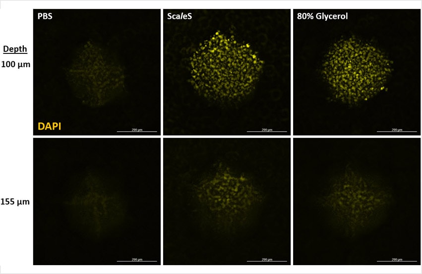
Many clearing agents and mounting media have been developed to help eliminate diffracted light by altering the refractive index of the sample so that they better match the refractive index of the microscope optics. Selecting the most appropriate clearing agent requires consideration of multiple factors, including: 1) sample thickness, 2) imaging depth, 3) fluorophore type, and 4) vessel format and material. Some clearing agents function by simply exchanging the primarily water-based intracellular environment with a liquid that has a better matched refractive index, such as glycerol.
Alternatively, some clearing agents improve sample clarity by extracting lipids (delipidation). Clearing agents that simply exchange the intracellular aqueous environment are minimally invasive and tend to favor smaller samples and those where fluorescent proteins are used. While clearing agents that delipidate may improve imaging depth to a greater degree, these agents typically employ harsh detergents and electrophoretic devises to draw lipids away from the sample. Some delipidating clearing agents use organic solvents, which may be incompatible with fluorescent protein-based samples. Additionally, organic solvent-based clearing agents may not be compatible with imaging vessels that use glass alternatives as the imaging window, such as cycloolefin. To that point, choosing the right interface between your sample and the microscope objective is crucial to achieving the best matched refractive index possible (Figure 1).
Most microscopes are designed based on how light passes and diffracts through a range of coverslip thicknesses made of glass, from about 170 nm to 2 mm, which can accounted for using the objective's correction collar (if it has one). Light passes through plastic differently, and imaging through such a material, regardless of its thickness, may compromise image quality.
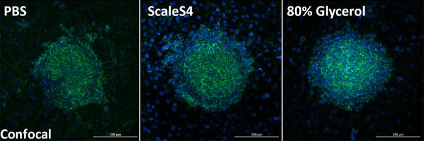
Figure 1. Comparison of clearing methods using adhered HT1080 Spheroids. HT1080 spheroids were formed by seeding 1000 cells into ultra low attachment (ULA) U-bottom 96-well plates (Corning Cat# 4520) and allowed to coalesce for 24 hours at 37°C. Spheroids were then transferred to clear, flat-bottom 96-well plates (Agilent Technologies Cat# 204626) (1 spheroid/well) and allowed to settle and establish adhesion at RT for 1 hr. Plates containing spheroids were then transferred back to the 37°C incubator to allow spheroids to attach to the culture area and spread overnight before fixing samples with 4% PFA for 30 min, then permeablizing with 0.5% for 1 hour. To achieve full penetration of dyes, spheroids were stained with Hoechst 34580 and Alexa Fluor 488-phalloidin overnight at 4°C. Spheroids were then washed, and PBS was either maintained, or exchanged for either the ScaleS4 (40% D-Sorbitol, 10% Glycerol, 4M Urea, 15% DMSO, 0.5% n-Propyl Gallate, 0.05% NaN3), or a homemade mounting medium (80% glycerol, 20 mM Tris, pH 8.0, 0.5% n-Propyl Gallate, 0.05% NaN3). Confocal z-stack images of fixed and stained HT1080 Spheroids were captured at 40X using the Cytation C10 Confocal Imaging Reader and projected as a single image.
Optical Sectioning

By removing contaminating out-of-focus light, confocal microscopy affords more resolved, thinner sections in the Z axis compared to widefield microscopy. To fully take advantage of this improvement, it is important to image a sufficient amount of sections such that a more accurate three dimensional render of the sample can be constructed. The Nyquist sampling theorem provide a mathematical foundation to determine the step sizes (and thus, sampling rate in the Z axis) to gain the best depiction of your sample possible. For microscopy, determining the Z step size that give the appropriate Nyquist sampling rate involves considering the numerical aperture (NA) of your objective, the emission wavelength of your fluorescent probe, and the refractive index of the interface between the objective and the coverslip. Plugging in these values into the equation will results in a step size that achieves Nyquist sampling. Collecting sections thinner than this is considered oversampling and can improve 3D rendering to a certain extent. Collecting Z-sections thicker than Nyquist is considered undersampling compared to what is possible with the microscope, and should be conducted judiciously. If the aspects of your sample do not require such rigorous sectioning, but the analysis or interpretation can still be improved with confocal microscopy, this is certainly something to consider. The more sectioning that occurs comes with the caveat of generating more data that will need to be managed.
Webinar
Cytation C10: An Affordable Confocal Imaging Reader for your Research Laboratory
Presenter: Chris Laucius, Ph.D, Product Manager in Development at BioTek Instruments
Cytation C10 Confocal Imaging Reader combines automated confocal and widefield microscopy with conventional multi-mode microplate reading in a compact, modular design. The spinning disk confocal module provides exquisite detail and resolution in a wide variety of sample types that benefit from confocality. High quality components are used for excellent image quality, at a lower price than similar systems.
Cytation C10 also includes widefield fluorescence, brightfield and phase contrast optics. Its variable bandwidth monochromator-based plate reading provides fluorophore specificity and sensitivity. The combination of confocal and widefield imaging with multi-mode detection will add efficiency to your lab workflows and help increase productivity. Cytation C10 is ready for any assay.
For Research Use Only. Not for use in diagnostic procedures.