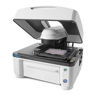
BioTek Lionheart FX 全自动智能成像分析系统
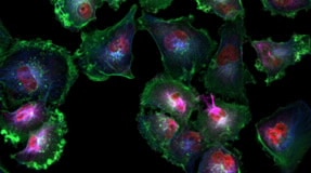
Lionheart FX 全自动智能成像分析系统是一款一体化系统,可用于广泛的成像工作流程。它可使用高达 60x 和 100x 的油镜,具有荧光、明场、彩色明场和相差成像模式,可大大扩展应用范围。
可选的环境控制盖可实现 40 °C 温控,并能有效控制环境 CO2/O2 的浓度。湿度控制仓可以优化长时间活细胞成像应用的环境条件,并且双自动进样器有助于进行快速的动力学分析。
- Automated Cell Imagers
Request a Quote
使用上方的下载按钮下载产品样本,或 索取关于 Lionheart FX 全自动智能成像分析系统的更多信息
Product Details
Features
- 体积小巧,可轻松安装在大多数标准实验室工作台上
- 图像采集无需外部仪器或专用暗室
- 宽场 (WFOV) 相机可对微孔板和切片进行快速自动化成像,使研究人员能够快速获得所需的答案
- Gen5 成像软件提供独特的全自动采集、处理、分析和发表工作流程
- 环境控制功能使 Lionheart FX 能够长时间对活细胞进行成像和分析
- 基于激光和基于软件的自动聚焦可在自动显微镜中采集清晰、对焦的图像
- Lionheart FX 载物台采用开放式设计,便于研究人员使用微流控设备
- Lionheart FX 的双加样器模块有助于在添加试剂后对快速细胞反应进行成像
Specifications
| 成像仪与微孔板检测仪选件 |
|
| 成像方法 |
|
| 成像模式 |
|
| 支持的样品容器 |
|
| 放大倍数范围 |
|
| 显微镜类型 |
|
| 环境控制 |
|
How It Works
Applications
Technology
自动识别目标区域
此过程与安捷伦自动化产品相结合,可对四张或更多张玻片实现无人值守玻片扫描,并且可与 Agilent BioTek Lionheart 全自动智能成像分析系统和 Agilent BioTek Cytation 成像多功能微孔板检测仪配合使用。
Learn More点计数
Agilent BioTek Gen5 软件中提供的点计数模块能够将计数的点数量和其他信息与原始一级 Mask 数据相关联,使这些值对于图像中最初鉴别出的每个目标是已知的。
Learn More4-Zone 温控和抗凝集功能
多款安捷伦单功能和多功能微孔板检测仪都具备用于温度敏感型应用(例如动力学分析)的温控功能。某些微孔板检测仪的培养箱使用带风扇的加热模块来循环热空气,为读数室和微孔板保温。
Learn MoreLiterature
- Key Literature
-
Download Literature
Download a product brochure or data sheet
- Brochures
- English
- 23 Nov 2021
- 72.95 KB
- Application Notes
- Article Reprints
- Posters
- Selection Guides
- Specifications
- Technical Overviews
Tools
Image Gallery
Click here to view our Image Gallery, including images captured with Agilent BioTek instruments.
Videos
Training & Events
Agilent Cell Analysis Solutions: Empowering scientific discovery and advancing groundbreaking therapeutics
Unlock the potential of cutting-edge Agilent Cell Analysis technologies in this insightful webinar.
Webinar/Training
Agilent BioTek Lionheart Technology: Driving physiological relevance in early drug discovery
This presentation focuses on the ability to build a comprehensive story from drug screening to histological confirmation through a combinatorial approach on a single platform.
Webinar/Training
A Practical Guide for 3D Cell Culture Systems and Optimizing Spheroid Imaging Assay - Part 2 of 2
Three-dimensional (3D) cell culture systems have become a well-established in vitro experimental approach to model human disease for a broad range of research applications.
Webinar/Training
Support
- FAQs
-
- Site Preparation Checklists
Didn’t find what you need? Contact our dedicated Cell Analysis support teams.
Related Products
Promotions