Key Takeaways
- Critical Role of Digital Pathology: Digital pathology, using whole-slide imaging (WSI) and artificial intelligence (AI), is transforming cancer diagnosis by improving precision, efficiency, and specialist collaboration. This is essential to meet the demands of precision medicine and complex testing.
- Challenges and Solutions: Despite advancements, issues like data quality, variability, AI explainability, and standardization need to be addressed for wider clinical adoption and effective integration into routine practice.
- Data Management and Storage: The large data volumes from digital pathology, especially high-resolution WSI files, require effective storage solutions. Cloud and hybrid storage approaches help manage these demands, ensuring long-term data preservation and accessibility.

Pathology plays a critical role in cancer care, encompassing the development of new treatments, diagnosis, staging, grading of disease, and clinical decision-making. While histopathological slides of tissue biopsies using hematoxylin and eosin (H&E) staining and immunohistochemical (IHC) staining remain central to this process (H&E staining provides a general view of tissue structure, while IHC staining identifies molecular markers), both are crucial for precision medicine testing, and the rise in testing1 is placing greater demands on pathology labs. Healthcare organizations globally are also grappling with resource shortages to address this challenge, mainly due to the declining number of medical professionals2 choosing pathology as a specialty. Additional challenges include increased testing rates, driven by more widespread screening programs and advancements in medical technology3, the rising incidence of cancer, and the growing complexity of testing.
Many research and clinical labs aim to enhance workflow efficiency and improve analytical quality by digitalizing processes to address the growing demand amid limited resources4. This approach leverages computational resources to reduce the burden on individual pathologists, enabling them to focus their expertise where it is most impactful. Following the evolutionary path of radiological and cardiological imaging, pathologists have embraced digital pathology and AI as a significant step forward in histopathology5. This also aims to enhance communication between specialists across the healthcare spectrum, both within pathology and beyond. The goal is to achieve faster, more consistent quality diagnoses.

Expanding role for digital pathology
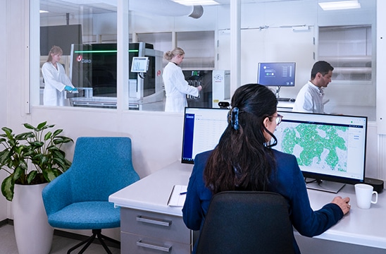
Central to the evolution of digital pathology is whole-slide imaging (WSI), which uses high-resolution scanners to capture and digitally stitch together microscopy images of tissue samples. Images can be taken at varying magnifications, offering insights from tissue morphology and immune invasion to nuclear identity and biomarker localization. Beyond its cartographic analog, WSI can provide 3-D insights by altering image focus along a tissue’s z-axis (z-stacking), which is essential for characterizing the tumor microenvironment (TME).
One key advantage of digitalization6 is that it facilitates remote consultation and telepathology, allowing pathologists to easily connect with colleagues for opinions on patient biopsies or enabling research collaborators to share and discuss experimental findings. It also helps centralize the analysis of samples taken at different locations within a healthcare network.
Physical slides may degrade over time due to environmental factors such as light and temperature or administrative challenges like mislabeling or breakage. In contrast, WSI digitally preserves them within an archive. This allows for analysis years after their original retrieval and, if desired, connection to other patient information, such as electronic health records (EHRs)7.
WSI offers pathologists a wealth of information, providing a richer picture of a tissue sample, but this abundance can also overwhelm the analytical process. For example, because WSI involves scanning an entire microscope slide to create a high-resolution digital image, this process increases the amount of data by providing more cells to analyze. This can complicate the analysis process for pathologists who need to focus on specific, clinically relevant regions of interest (ROIs) of the tissue sample. This is where advances in AI offer possible solutions.
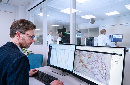
Trained digital pathology algorithms such as those from Visiopharm can automate various steps in the analysis process, such as identifying different cell types and nuclear morphologies within a tissue, defining regions of normal vs abnormal tissue, highlighting immune infiltration, indicating the location and intensity of immunohistochemical stains and biomarkers, and enumerating mitotic status and other features that inform a pathologist’s interpretation of a slide. Measuring these key characteristics enables pathologists to concentrate on a more in-depth, qualitative analysis of regions of interest (ROIs). In this way, AI doesn’t replace the pathologist but facilitates their efforts, creating what has been described as augmented intelligence.
Navigating the challenges of AI in digital pathology
Although digital pathology platforms have made significant inroads in the clinic over the last decade, much still needs to be accomplished before many AI algorithms are available outside the research wing. As with any large-scale computational effort, data quality, volume, and variability continue to challenge the evolution of these algorithms from the idealized laboratory setting to real-world clinical application8.
The challenge of explainability in AI:Explainability also remains challenging with AI, as clinicians and regulatory agencies want to know how an algorithm reaches its conclusions. AI platforms are sometimes called “black boxes” to anyone unfamiliar with computational science. For clinical use, explainability helps the pathologist determine how much they wish to rely on the platform’s recommendation, particularly in cases where their opinions differ. User interface features can facilitate this by highlighting the parameters that led the AI system to a decision.
Navigating regulatory approval for AI algorithms:Regarding regulatory approval of AI algorithms, the challenge presents itself in two forms. When the AI platform effectively confirms and supports a widely accepted and validated diagnostic method, the path to regulatory approval should be smoother, and the question is more likely to center on how the platform improves on what already exists. Part of the power of AI, however, is its capacity to identify and correlate diagnostic and prognostic patterns and features not previously identified. In such a situation, the approval process will demand not only validation of the platform but also the clinical soundness of the identified patterns and features9.
The need for standardization in digital pathology:Standardization will also be critical to the broader adoption of digital pathology. Unlike radiological platforms, which conform to the digital imaging and communications in medicine (DICOM) format, slide scanner manufacturers produce image data in various formats, adding complexity to routine diagnosis. The move toward vendor-agnostic standards would provide a common language, facilitating efforts to connect digital pathology data to other clinical systems, such as radiology picture archiving and communications systems (PACS), laboratory information systems (LIS), and EHRs.
Challenges in developing AI algorithm training and validation data:Even within the research space, the need for standards complicates the development of AI algorithm training and validation data, which may arise from multiple sources. For example, platforms developed with data from just one institution might not work as effectively with data from other institutions due to unique characteristics in the original data. This could lead to less accurate results when the platform is used more broadly. Broad agreement on standards and the types of metadata associated with WSIs would help address these challenges10.
Storage strategies for digital pathology data
The opportunities for digital pathology to facilitate the sharing, analysis, and storage of pathology images are apparent, implementation of the practices is not without significant challenges, particularly regarding image storage. With high-resolution WSI files approaching two to four gigabytes each, it is easy to imagine a typical digital pathology practice generating up to a petabyte of data annually. These IT demands are further exacerbated by long-term storage requirements that can see images preserved for up to 20 years. The costs of storing such large and growing volumes of data are substantial and include not only the expansion and maintenance of the physical storage media but also the maintenance and security of the data.
Optimizing storage with cloud technology:Cloud storage solutions offer scalable storage options that can grow with the increasing data volumes, providing easier access to data from multiple locations and potentially being more cost-effective with a pay-as-you-go model. The distributed uploading and accessing of large WSI files places heavy demands on bandwidth and internet connection fidelity. With fewer in-house infrastructure demands, Cloud storage would likely benefit smaller laboratories that may wish to dedicate their limited financial and organizational resources to demands other than extensive server capacity11.
A hybrid storage approach:Larger, higher-traffic laboratories, however, may be more interested in a hybrid approach that combines on-premises storage for frequently accessed data with cloud storage for long-term archiving. This approach optimizes infrastructure costs while also facilitating pathologist access to critical, clinical decision-making data. The hybrid approach also offers enhanced security by keeping sensitive data on-premises while leveraging the cloud for less critical data, providing the flexibility to balance performance, cost, and security based on the laboratory's specific needs.
The potential to revolutionize cancer diagnosis
Integrating digital pathology holds immense potential for pathologists to revolutionize cancer diagnosis and aid in selecting appropriate treatments. While significant progress has been made with WSI and AI algorithms, challenges such as data quality, variability, and explainability remain. Efforts to standardize data formats and validate AI platforms across diverse real-world conditions continue and are crucial for broader clinical adoption. As these technologies evolve, they promise to enhance diagnostic accuracy and streamline workflows by providing pathologists with powerful tools to manage the growing complexity of cancer care.
Unless otherwise stated all products are Research Use Only, not for use in clinical diagnostic procedures. The mentioned digital pathology solutions are not available in all countries and may have different regulatory statuses. Please check with your local Agilent representative for availability in your country of use.
- https://www.avalonhcs.com/2024-lab-trend-report/
- https://thepathologist.com/issues/2023/articles/feb/constant-demand-patchy-supply/
- https://www.ons.org/publications-research/voice/advocacy/12-2024/5-common-cancers-prevention-and-screening-save-more
- https://link.springer.com/chapter/10.1007/978-3-031-82971-0_6
- https://medicine.iu.edu/magazine/issues/winter-2025/how-ai-is-revolutionizing-pathology
- https://www.pathologynews.com/digital-pathology/telepathology-101-patient-care-prospects-and-challenges/
- https://meridian.allenpress.com/aplm/article/143/2/222/64743/a-practical-guide-to-whole-slide-imaging-a-white
- https://www.mayoclinicplatform.org/2021/07/30/the-future-belongs-to-digital-pathology/
- https://www.thomsonreuters.com/en-us/posts/technology/ai-regulatory-challenge/
- https://www.frontiersin.org/journals/digital-health/articles/10.3389/fdgth.2025.1567624/abstract
- https://hal.science/hal-02889947v1